含谷胱甘肽透明质酸水光联合 1064nm Nd:YAG 激光及口服氨甲环酸治疗黄褐斑:一项前瞻性对照研究(Medicine, 2026)
2026年10月27日35 min read
Medicine
Medicine
结论与实践要点
该研究在 50 例中国女性表皮型黄褐斑患者中比较了「激光 + 口服氨甲环酸」基础上是否叠加皮内 HA-谷胱甘肽水光(HA-GSH)的疗效。最终每组 18 例完成、每组脱落 7 例(均与不良事件无关)。以改良黄褐斑面积和严重度指数(mMASI)为主要指标,校正基线后 A 组(加 HA-GSH)在全部随访点的平均差值较 B 组多 0.72 分(95%CI 0.46–0.98,P<.001),且自第 3 次治疗起差异显现;治疗结束(6 月)时 A 组 mMASI 2.50 显著低于 B 组 3.39(P<.001),停药 2 月后优势仍保持(2.06 vs 2.67,P=.003)。两组均仅出现短暂红斑、轻度肿胀与淤青,未见严重或色素异常不良事件,提示联合方案耐受性较好。
临床落地需注意边界:本研究为单中心、未设安慰剂/盲法(设备治疗难以盲法)、且 2 月随访尚不足以评估黄褐斑这一慢性易复发疾病的长期复发情况;A 组满意度数值更高但未达统计学显著。叠加 HA-GSH 可作为激光-氨甲环酸方案的辅助增效选择,但其长期维持获益、最佳注射方案与成本效益仍需在更大样本、更长随访的试验中确认。此外口服氨甲环酸应关注血栓等禁忌与用药监护。
Melasma is a chronic, acquired facial hyperpigmentation disorder with multifactorial pathogenesis and frequent relapse. Low-fluence 1064-nm Q-switched neodymium-doped yttrium aluminum garnet (Nd:YAG) (LQSNY) laser with oral tranexamic acid (TA) is widely used, but responses are often incomplete and short-lived. glutathione (GSH), an antioxidant that inhibits melanogenesis, may improve outcomes when delivered intradermally with a hyaluronic acid (HA) carrier. We assessed whether adding an HA-based GSH skin booster (HA-GSH) to this regimen enhances efficacy and durability. Fifty Chinese women with epidermal melasma (Fitzpatrick III–IV) were randomized 1:1 to 6 monthly LQSNY sessions plus oral TA (250 mg daily), with (Group A) or without (Group B) intradermal HA-GSH. The primary outcome was change in modified melasma area and severity index (mMASI) at 6 months and at 2-month posttreatment follow-up. Secondary outcomes were patient satisfaction (short assessment of patient satisfaction) and safety. Sample size was determined a priori. mMASI was analyzed using a linear mixed-effects model with baseline mMASI as a covariate and Benjamini–Hochberg adjustment for visit-level comparisons. Thirty-six patients completed the study (18 per group); baseline mMASI was comparable (5.61 ± 1.29 vs 5.56 ± 1.20; P = .89). Fourteen participants withdrew (seven per group), none due to adverse events. Group A showed significantly greater mMASI reduction from the third session onward. Adjusted for baseline, the mean difference across all post-baseline visits was 0.72 points (95% confidence interval 0.46–0.98; P < .001) favoring Group A, evident at the first posttreatment assessment. At 6 months, mean mMASI was lower in Group A (2.50 ± 0.62 vs 3.39 ± 0.70; P < .001), an advantage maintained at follow-up (2.06 ± 0.54 vs 2.67 ± 0.59; P = .003). More Group A patients were “very satisfied” (28% vs 11%), though not significantly ( P = .31). Both regimens were well tolerated; only transient erythema, mild swelling, and bruising occurred, with no serious or pigmentary adverse events. Adding intradermal HA-GSH to LQSNY laser and oral TA produced earlier, greater, and more durable melasma improvement with high satisfaction and a favorable safety profile, supporting HA-GSH as a promising adjuvant pending confirmation in larger, longer trials.
Keywords: combination therapy、glutathione、hyaluronic acid skin booster、melasma、mMASI、Q-switched Nd:YAG laser、tranexamic acid
Melasma is a common acquired disorder of hyperpigmentation characterized by symmetrical dark patches on sun-exposed areas of the skin, most often the face. It is also referred to as “chloasma” or the “butterfly mask.” [ 1 ] The condition is most prevalent in women with Fitzpatrick skin types III to V, particularly within Asian populations. Although benign, melasma frequently causes psychological distress and significantly reduces quality of life. [ 2 ]
The pathogenesis of melasma is multifactorial, involving ultraviolet (UV) radiation, hormonal influences, oxidative stress, and genetic predisposition. Available treatments include topical depigmenting agents such as hydroquinone and azelaic acid, oral tranexamic acid (TA), chemical peels, and laser-based therapies. [ 3 ] However, monotherapies usually yield incomplete or temporary improvement, and relapse is common.
The low-fluence 1064-nm Q-switched neodymium-doped yttrium aluminum garnet (Nd:YAG) (QSNY) (LQSNY 1064-nm) laser is widely used because it can selectively target melanin granules in melanocytes while sparing surrounding tissues, thereby minimizing scarring. [ 4 ] Enhanced outcomes have been reported when this laser is combined with adjuvant therapies, including TA, [ 5 ] azelaic acid, [ 6 ] and kojic acid. [ 7 ]
Glutathione (GSH), a tripeptide antioxidant naturally present in human tissues, has also been investigated as a potential therapy for melasma. It decreases tyrosinase activity, shifts melanin synthesis from eumelanin to pheomelanin, and scavenges reactive oxygen species generated by UV radiation. [ 8 ] By contrast, TA reduces melanogenesis primarily through inhibition of plasminogen activation and has demonstrated efficacy in multiple controlled trials. [ 9 ]
The present study prospectively evaluated the efficacy and safety of LQSNY 1064-nm laser combined with oral TA, with or without intradermal hyaluronic acid (HA)–based skin booster containing GSH (HA-GSH). The modified melasma area and severity index (mMASI) was used as the primary outcome measure.
This study was conducted in accordance with the Declaration of Helsinki (1975) and approved by the institutional review board of 3201 Hospital. All participants provided written informed consent after being informed of the study procedures, potential risks, benefits, and complications.
Fifty healthy Chinese women with clinically diagnosed melasma (Fitzpatrick skin types III or IV), aged 24 to 51 years, were enrolled. Exclusion criteria included age < 18 years, pregnancy or lactation, dermal or mixed-type melasma, a history of keloids, photosensitivity, bleeding disorders, chronic liver disease, autoimmune or thyroid disease, active asthma, or recurrent herpes simplex infection. Patients with active facial infections or inflammatory skin conditions were excluded, as were those with recent exposure to hormonal therapies (e.g., oral contraceptives), anticoagulants, antiepileptics, or chemotherapeutic agents. Prior treatment with laser/light therapy within 6 months, oral or topical retinoids within 12 months, topical depigmenting agents within 4 weeks to 3 months, or chemical peels and TA within 3 months also led to exclusion. Hypersensitivity to any study medication component was an additional criterion.
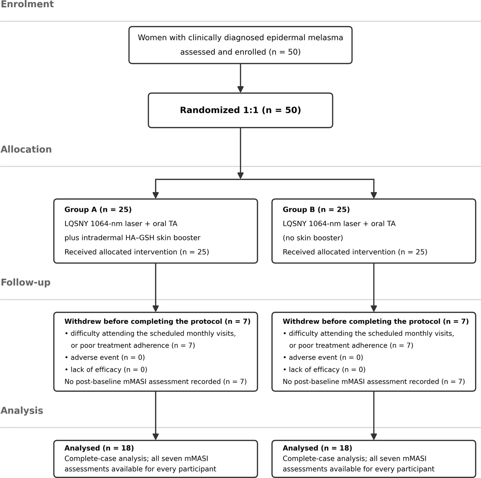
Patients were randomized in a 1:1 ratio using a computer-generated sequence. Allocation was concealed with sealed envelopes coded as group A or group B. Group A received LQSNY 1064-nm laser combined with oral TA plus HA-GSH. Group B received LQSNY 1064-nm laser with oral TA alone. (Fig. 1 )
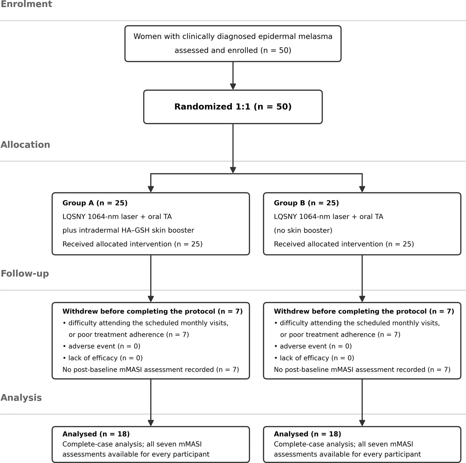
Figure 1. Study flowchart diagram (CONSORT-style flow diagram showing the numbers screened, randomized, withdrawn from each group with reasons for withdrawal, and analyzed). CONSORT = Consolidated Standards of Reporting Trials.研究流程图(CONSORT 风格),展示筛选、随机分组、各组退出例数及原因与最终分析人数。
The sample size was determined a priori for the primary endpoint, the mMASI score at the end of the 6-month treatment course. Drawing on the magnitude of benefit reported when GSH is added to a procedural intervention for facial melasma, we assumed a between-group difference in mMASI of 1.2 points with a common standard deviation of 1.35 points, corresponding to a standardized effect size (Cohen d ) of 0.89. For a 2-sided independent-samples t -test with α = 0.05 and 80% power, 20 participants per group were required. Allowing for an anticipated attrition rate of approximately 20%, we planned to enroll 25 participants per group, for a total of 50. Calculations were performed with G*Power version 3.1.9.7 (Heinrich Heine University).
Because 36 participants (18 per group) ultimately completed the study, a post hoc sensitivity analysis of precision was undertaken. With 18 participants per group, α = 0.05 (2 sided) and 80% power, the smallest detectable standardized difference was d = 0.93, equivalent to approximately 0.62 mMASI points at the observed variability of the primary endpoint. The difference actually observed at the end of treatment corresponded to d = 1.35, and the achieved power for that comparison was 98%. The study therefore retained adequate power for the primary endpoint, although power for smaller effects and for the secondary patient satisfaction outcome was limited.
Thirty-six of the 50 patients (aged 30–44 years; median, 36) completed the study. Fourteen participants (28%) withdrew before completing the protocol, 7 from each group. In every case, the reason was logistical, difficulty attending the scheduled monthly follow-up visits or poor adherence to the treatment protocol. No withdrawal was attributable to an adverse event, to lack of efficacy, or to a protocol-defined safety criterion (Fig. 1 ). Because these participants left the study before any post-baseline mMASI assessment was recorded, no on-treatment efficacy data were available for them, and the efficacy analysis was therefore a complete-case analysis of the 36 participants who completed the protocol; the implications are addressed in the statistical analysis and in the Discussion. A comprehensive medical history, family history, and dermatological assessment were performed at baseline, including Fitzpatrick phototype and melasma clinical subtype. Melasma clinical subtype was determined at baseline by an experienced dermatologist using clinical examination combined with Wood lamp (365 nm) examination: lesions showing enhancement of pigment contrast under Wood light were classified as epidermal type, those without enhancement as dermal type, and those showing both features as mixed type. Only epidermal-type melasma was eligible for inclusion, whereas dermal- and mixed-type cases were excluded.
All patients underwent facial cleansing before laser therapy. Both sides of the face were treated with 6 monthly sessions of LQSNY 1064-nm laser. Parameters included a 6-mm spot size, collimated homogeneous flat-top beam profile, fluence of 2.2 to 2.6 J/cm 2 , and frequency of 10 Hz. The clinical endpoint was immediate pigment lightening or the appearance of mild erythema without petechiae. Concurrently, oral TA (250 mg once daily) was administered for 6 months.
In group A, patients also received intradermal injections of a compounded solution consisting of 0.5 mL of injectable GSH (0.6 g dissolved in 2 mL sodium chloride injection) mixed with 2 mL of a HA-based skin booster, yielding a total volume of 2.5 mL. A topical anesthetic cream containing 10% lidocaine was applied for 20 to 30 minutes under occlusion before each session. The solution was injected into the deep dermis or superficial subcutaneous layer of the entire face, with particular focus on melasma lesions, using 25-G cannulas. Six monthly sessions were performed in parallel with laser treatments.
posttreatment instructions included avoiding facial cleansing for 24 hours and refraining from additional melasma therapies during the study period. Patients were advised to strictly limit sun exposure and to apply broad-spectrum sunscreen with SPF ≥ 50 daily. Temporary erythema, swelling, bruising were anticipated and typically resolved within 2 to 3 days.
Evaluation of treatment efficacy was performed using the mMASI, calculated for the face at baseline, before each treatment session (monthly, for a total of 6 sessions over 6 months), and again 2 months after completion of treatment. Patient satisfaction was assessed with the validated seven-item short assessment of patient satisfaction scale. [ 8 ] Short assessment of patient satisfaction scores were interpreted as follows: 0 to 10, very dissatisfied; 11 to 18, dissatisfied; 19 to 26, satisfied; 27 to 28, very satisfied. At each visit, adverse events related to either the medication (e.g., nausea, vomiting, heartburn, thromboembolic events) or the laser therapy (e.g., persistent erythema, post-inflammatory hyperpigmentation, or hypopigmentation) were recorded. Standardized clinical photographs were obtained at every visit under fixed conditions (identical camera, lighting, distance, and patient positioning). To minimize observer bias, all de-identified baseline and final photographs were independently rescored by a second dermatologist who was blinded to group assignment and to assessment time point and who evaluated the images in randomized order; the blinded scores were consistent with those of the treating dermatologist.
Data were analyzed using International Business Machines Statistical Package for the Social Sciences Statistics version 20.0 (IBM Corp.). Qualitative variables were expressed as counts and percentages, and group differences were assessed using the chi-square test. Quantitative data were presented as mean ± standard deviation for normally distributed variables, or as median with range for non-normally distributed variables. Between-group comparisons were performed with independent-samples t -tests for normally distributed data. Correlations between continuous variables were assessed using Pearson correlation coefficient. Distributional assumptions were examined with the Shapiro–Wilk and Levene tests; because mMASI was recorded to the nearest whole point and departed from normality at several visits, all between-group comparisons were repeated with the Mann–Whitney U test as a sensitivity analysis. Fisher exact test replaced the chi-square test whenever an expected cell count was below 5. A 2-sided P value < .05 was considered statistically significant.
Longitudinal mMASI data were analyzed with a linear mixed-effects model for repeated measures, which accounts for the within-participant correlation of serial measurements. The model included treatment group, visit (entered as a categorical factor), the group × visit interaction, and the baseline mMASI score as fixed effects, with an unstructured within-participant covariance matrix estimated by restricted maximum likelihood and Kenward–Roger degrees of freedom; entering the baseline score as a covariate rather than as an outcome is the recommended approach for randomized trials and improves precision. The overall treatment effect was assessed as the group effect across the 6 post-baseline visits and summarized as the adjusted mean difference averaged over those visits, with estimated marginal means and 95% confidence intervals derived at each visit. The group × visit interaction was examined to determine whether the magnitude of the treatment effect changed over time. For completeness, the unadjusted group × time interaction across all seven assessments was also tested; sphericity was evaluated with Mauchly test and, because it was violated, the Greenhouse–Geisser correction was applied to the univariate analysis alongside the corresponding multivariate test.
Between-group comparisons at individual visits were regarded as secondary and were adjusted for multiplicity across the 6 post-baseline assessments using the Benjamini–Hochberg false-discovery-rate procedure; both unadjusted and adjusted P values are reported. No adjustment was applied to the overall treatment effect, which constituted a single test, or to the analyses of patient satisfaction and safety, which were descriptive. Because no post-baseline mMASI assessment was available for any of the 14 participants who withdrew, the efficacy analysis was necessarily a complete-case analysis of the 36 participants who completed the protocol, within which no observations were missing; no imputation was performed for the primary analysis, which accordingly rests on the assumption that the withdrawals were missing completely at random. To quantify the potential influence of the withdrawals, a pattern-mixture sensitivity analysis was undertaken in which plausible outcomes were assigned to the withdrawn participants under a range of assumptions (including a jump-to-reference scenario in which participants who withdrew from group A were assumed to have responded no better than group B) together with a tipping-point analysis identifying the assumed outcome at which the between-group difference would cease to be statistically significant.
Thirty-six patients completed the study (18 in each group). Attrition was balanced across arms (7 of 25 in each group; 28%). Baseline demographic and clinical characteristics were well balanced between the 2 groups. There were no significant differences in age, marital or reproductive status, residence, occupation, comorbidities, Fitzpatrick skin type, or disease duration. Clinical subtype distribution, disease course, history of previous treatment, family history of melasma, pregnancy-related exacerbation, and daily sun exposure were also comparable (all P > .05). (Fig. 1 ; Table 1 ).
Table 1 Baseline demographic and clinical characteristics.
| Characteristic | Group A (n = 18) | Group B (n = 18) | Test statistic | P |
|---|---|---|---|---|
| Age (yrs) | 36.33 ± 4.39 | 36.17 ± 4.31 | t = +0.11 | .909 |
| Disease duration (yrs) | 7.44 ± 2.12 | 6.33 ± 2.40 | t = +1.47 | .150 |
| Marital status, married/ single | 14/ 4 | 15/ 3 | Fisher exact | 1.000 |
| History of pregnancy, yes/ no | 13/ 5 | 14/ 4 | Fisher exact | 1.000 |
| Residence, rural/ urban | 12/ 6 | 14/ 4 | Fisher exact | .711 |
| Occupation, housewife/ employed | 14/ 4 | 15/ 3 | Fisher exact | 1.000 |
| Underlying disease, HTN/ DM/ thyroid/ none | 1/ 2/ 0/ 15 | 0/ 1/ 1/ 16 | χ 2 = 2.37, df = 3, sparse | .500 |
| Fitzpatrick skin type, III/ IV | 14/ 4 | 15/ 3 | Fisher exact | 1.000 |
| Clinical type, centrofacial/ malar/ mandibular | 9/ 1/ 8 | 10/ 1/ 7 | χ 2 = 0.12, df = 2, sparse | .942 |
| Disease course, stationary/ recurrent/ progressive | 10/ 2/ 6 | 12/ 1/ 5 | χ 2 = 0.61, df = 2, sparse | .739 |
| Previous treatment, yes/ no | 15/ 3 | 13/ 5 | Fisher exact | .691 |
| Family history of melasma, yes/ no | 6/ 12 | 5/ 13 | Fisher exact | 1.000 |
| Exacerbation during pregnancy, yes/ no | 12/ 6 | 13/ 5 | Fisher exact | 1.000 |
| Daily sun exposure, < 3 h/ ≥ 3 h | 12/ 6 | 10/ 8 | Fisher exact | .733 |
At baseline, mean mMASI scores were similar between group A (5.61 ± 1.29) and group B (5.56 ± 1.20; P = .89). Progressive improvement was observed in both groups during treatment. Before the third session, group A demonstrated significantly lower scores than group B (4.00 ± 0.84 vs 4.61 ± 0.85; P = .04). This trend continued through subsequent sessions, with group A showing consistently greater reductions (fourth session: 3.61 ± 0.78 vs 4.39 ± 0.92, P = .01; fifth session: 2.94 ± 0.73 vs 3.78 ± 0.73, P = .002; sixth session: 2.50 ± 0.62 vs 3.39 ± 0.70, P < .001). At the 2-month posttreatment follow-up (month 7), both groups maintained improvement; however, group A continued to show superior outcomes (2.06 ± 0.54 vs 2.67 ± 0.59; P = .003) (Table 2 , Fig. 2 ).
Table 2 mMASI over the treatment course.
| Time point | Group A (n = 18) | Group B (n = 18) | Difference (95% CI) | P | P (adj.) | P (M–W) | d |
|---|---|---|---|---|---|---|---|
| Baseline | 5.61 ± 1.29 | 5.56 ± 1.20 | 0.06 (−0.79–0.90) | .894 | - | .882 | 0.04 |
| Before session 2 | 4.67 ± 1.14 | 5.11 ± 1.13 | −0.44 (−1.21–0.32) | .248 | .248 | .319 | −0.39 |
| Before session 3 | 4.00 ± 0.84 | 4.61 ± 0.85 | −0.61 (−1.18–− 0.04) | .037 | .045 | .061 | −0.72 |
| Before session 4 | 3.61 ± 0.78 | 4.39 ± 0.92 | −0.78 (−1.35–− 0.20) | .010 | .014 | .015 | −0.92 |
| Before session 5 | 2.94 ± 0.73 | 3.78 ± 0.73 | −0.83 (−1.33–− 0.34) | .002 | .005 | .004 | −1.14 |
| Before session 6 | 2.50 ± 0.62 | 3.39 ± 0.70 | −0.89 (−1.34–− 0.44) | < .001 | .002 | < .001 | −1.35 |
| 2-month follow-up | 2.06 ± 0.54 | 2.67 ± 0.59 | −0.61 (−1.00–− 0.23) | .003 | .005 | .004 | −1.08 |
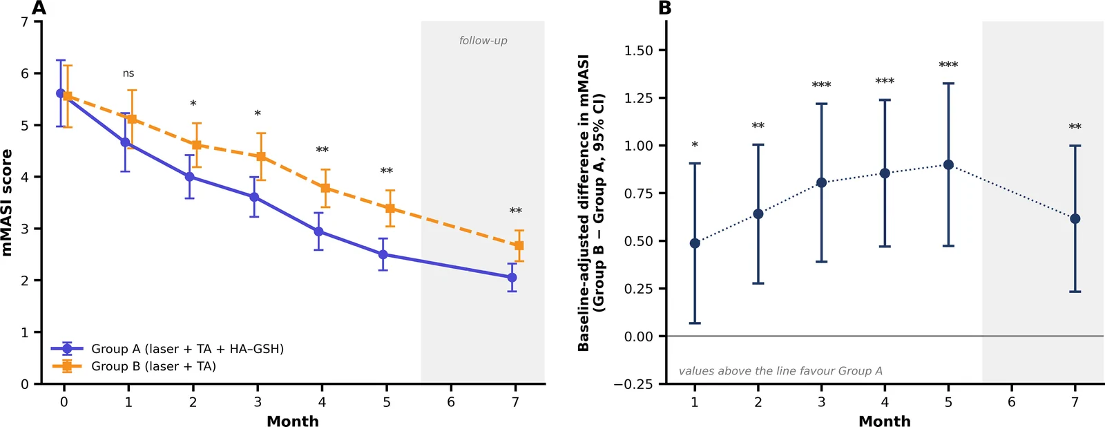
Figure 2. mMASI score between the 2 groups over sessions. mMASI = modified melasma area and severity index.两组 mMASI 评分随治疗次数变化曲线,直观显示 A 组(联合 HA-GSH)自中期起下降更明显、停药后维持更优。In the mixed-effects model for repeated measures, mMASI improved highly significantly over time in both groups (F(6, 29) = 42.2, P < .001; Table 3 ). After adjustment for the baseline score, the overall treatment effect across the 6 post-baseline visits was significant (Wilks’ λ = 0.499; F [6, 28 = 4.69, P = .002), corresponding to a mean adjusted difference of 0.72 mMASI points (95% CI 0.46–0.98; P < .001) in favor of group A. The adjusted between-group difference df avored group A at every post-baseline visit, including the first (before session 2: 0.49, 95% CI 0.07–0.91, adjusted P = .024; before session 3: 0.64, 95% CI 0.28–1.00, adjusted P = .002; before session 4: 0.81, 95% CI 0.39–1.22, adjusted P < .001; before session 5: 0.85, 95% CI 0.47–1.24, adjusted P < .001; before session 6: 0.90, 95% CI 0.47–1.33, adjusted P < .001; 2-month follow-up: 0.62, 95% CI 0.23–1.00, adjusted P = .003; Table 4 ). The magnitude of this effect did not change significantly across the treatment course (group × visit interaction, F [5, 29] = 0.75, P = .59), indicating an early benefit that was subsequently maintained rather than 1 that widened progressively.
Table 3 Linear mixed-effects model for repeated measures.
| Effect | Test statistic | P | Interpretation |
|---|---|---|---|
| Unadjusted analysis of all 7 assessments | |||
| Time (within-subject main effect) | F (6, 29) = 42.22 | < .001 | mMASI improved in both groups |
| Group (between-subject main effect) | F (1, 34) = 6.49 | .016 | Overall level differs between arms |
| Group × time, Greenhouse–Geisser corrected | F (2.48, 84.4) = 2.53 | .074 | Not significant |
| Group × time, multivariate | F (6, 29) = 1.70 | .158 | Not significant |
| Mauchly test of sphericity | χ 2 (20) = 108.4, ε = 0.414 | < .001 | Sphericity violated |
| Baseline-adjusted analysis of the 6 post-baseline visits | |||
| Group effect (overall treatment effect) | Wilks’ λ = 0.499; F (6, 28) = 4.69 | .002 | Significant treatment effect |
| Adjusted mean difference across visits | 0.72 (95% CI 0.46–0.98) | < .001 | In favor of Group A |
| Group × visit interaction | F (5, 29) = 0.75 | .594 | Effect constant over time |
| Linear trend of the treatment effect | t (33) = 1.43 | .163 | No progressive widening |
| Variance components (random-intercept parameterization) | |||
| Between-participant/ residual variance | σ 2 = 0.426/ 0.369 | - | ICC = 0.54 |
Table 4 Baseline-adjusted between-group differences in mMASI.
| Visit | Adjusted difference | 95% CI | P | P (adjusted) |
|---|---|---|---|---|
| Before session 2 | −0.49 | −0.91–− 0.07 | .024 | .024 |
| Before session 3 | −0.64 | −1.00–− 0.28 | .001 | .002 |
| Before session 4 | −0.80 | −1.22–− 0.39 | < .001 | < .001 |
| Before session 5 | −0.85 | −1.24–− 0.47 | < .001 | < .001 |
| Before session 6 | −0.90 | −1.33–− 0.47 | < .001 | < .001 |
| 2-month follow-up | −0.62 | −1.00–− 0.23 | .003 | .003 |
| Averaged across the 6 post-baseline visits | −0.72 | −0.98–− 0.45 | < .001 | - |
In the unadjusted analysis of all 7 assessments, Mauchly test indicated a marked departure from sphericity ( P < .001; Greenhouse–Geisser ε = 0.41). After correction, the group × time interaction did not reach statistical significance ( F [2.48, 84.4] = 2.53, P = .07; corresponding multivariate test F [6, 29] = 1.70, P = .16), whereas the main effect of group was significant ( F [1, 34] = 6.49, P = .016). The unadjusted between-group comparisons at individual visits remained significant from the third session onward after Benjamini–Hochberg adjustment (before session 3, adjusted P = .045; before session 4, adjusted P = .014; before session 5, adjusted P = .005; before session 6, adjusted P = .002; 2-month follow-up, adjusted P = .006). nonparametric Mann–Whitney comparisons gave the same pattern except before the third session, where the difference was of borderline significance ( P = .06) (Table 2 , Table 3 ).
Two pattern-mixture sensitivity analyses were used to gauge the influence of the 14 withdrawals on the primary endpoint. When participants who withdrew from group A were assumed to have responded no better than those in group B, the between-group difference at the end of treatment remained significant (0.64, 95% CI 0.17–1.10; P = .009). The tipping-point analysis indicated that the difference would cease to be significant only if the seven participants who withdrew from group A were assumed to have reached a mean mMASI of approximately 3.9 or higher, a poorer outcome than that actually achieved by the group B completers (3.39) and close to no improvement from baseline ( Table S1 , Supplemental Digital Content 1).
Within-group analysis revealed significant reductions in mMASI scores from baseline to month 7 in both groups (group A: from 5.61 ± 1.29 to 2.06 ± 0.54, mean reduction 3.56 ± 1.42, P < .001; group B: from 5.56 ± 1.20 to 2.67 ± 0.59, mean reduction 2.89 ± 1.08, P < .001). The absolute reduction was numerically larger in group A, but this raw change-score comparison did not reach statistical significance (mean difference 0.67 points, 95% CI −0.19–1.52; P = .12; Mann–Whitney P = .17), a comparison whose power is limited by the weak correlation between baseline and final scores in this cohort (group A, r = −0.05). When expressed as a proportion of the baseline score, the reduction was significantly greater in group A both at the end of treatment (53.1 ± 16.0% vs 37.8 ± 11.3%; P = .002) and at the 2-month follow-up (61.2 ± 12.8% vs 50.7 ± 12.1%; P = .016), consistent with the baseline-adjusted analysis reported above (Table 5 ).
Table 5 Change in mMASI from baseline to the 2-month follow-up.
| Baseline | Follow-up | Absolute reduction (95% CI) | Reduction (%) | P | |
|---|---|---|---|---|---|
| Group A (n = 18) | 5.61 ± 1.29 | 2.06 ± 0.54 | 3.56 ± 1.42 | 61.2 ± 12.8 | < .001 |
| Group B (n = 18) | 5.56 ± 1.20 | 2.67 ± 0.59 | 2.89 ± 1.08 | 50.7 ± 12.1 | < .001 |
| Between-group, absolute reduction | 0.67 (−0.19–1.52) | .123 | |||
| Between-group, proportional reduction at follow-up | 61.2 vs 50.7 | .016 | |||
| Between-group, proportional reduction at end of treatment | 53.1 vs 37.8 | .002 |
Patient satisfaction findings were numerically consistent with the clinical outcomes (Table 6 , Fig. 3 , Fig. 4 ). A higher proportion of patients in group A reported being “very satisfied” compared with group B (28% vs 11%). Most patients in both groups were “satisfied” (67% in group A; 72% in group B). Dissatisfaction was reported by 5% of patients in group A and 17% in group B. Although numerically more patients in group A were very satisfied, the distribution of satisfaction categories did not differ significantly between the groups ( χ 2 = 2.33, df = 2, P = .31). The study was not powered for this patient-reported outcome, and the satisfaction data should therefore be regarded as descriptive.
Table 6 Patient satisfaction at the end of the study (SAPS).
| Satisfaction category | Group A (n = 18) | Group B (n = 18) | SAPS score range |
|---|---|---|---|
| Very dissatisfied | 0 (0.0) | 0 (0.0) | 0–10 |
| Dissatisfied | 1 (5.6) | 3 (16.7) | 11–18 |
| Satisfied | 12 (66.7) | 13 (72.2) | 19–26 |
| Very satisfied | 5 (27.8) | 2 (11.1) | 27–28 |
| Total | 18 (100) | 18 (100) | - |
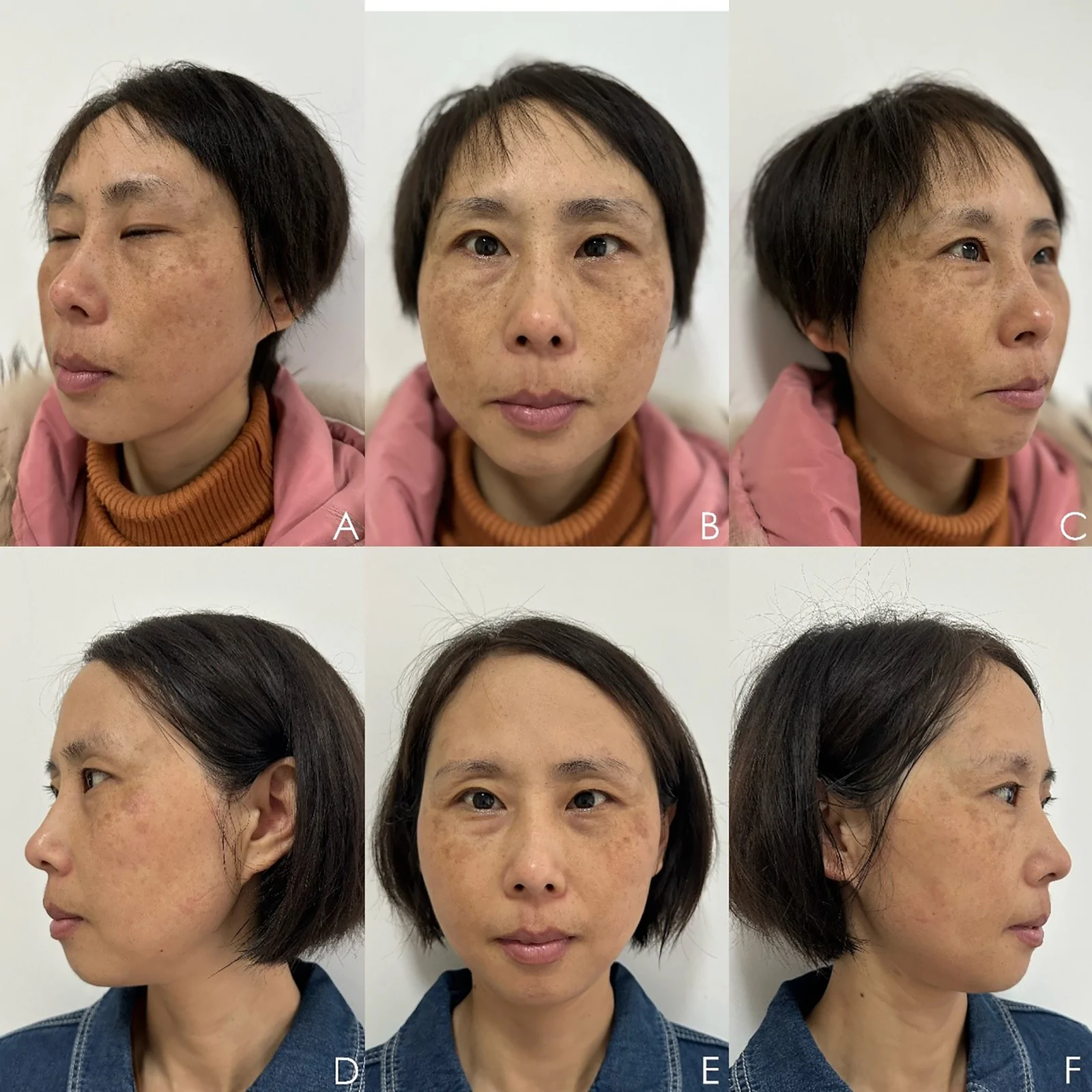
Figure 3. Baseline (A–C) and 2-month posttreatment (D–F) images of a Group A participant receiving 6 sessions of low-fluence 1064-nm Nd:YAG laser combined with oral tranexamic acid and intradermal glutathione/hyaluronic acid. Marked reduction of centrofacial pigmentation with improvement in skin clarity and overall complexion was observed. Nd:YAG = neodymium-doped yttrium aluminum garnet.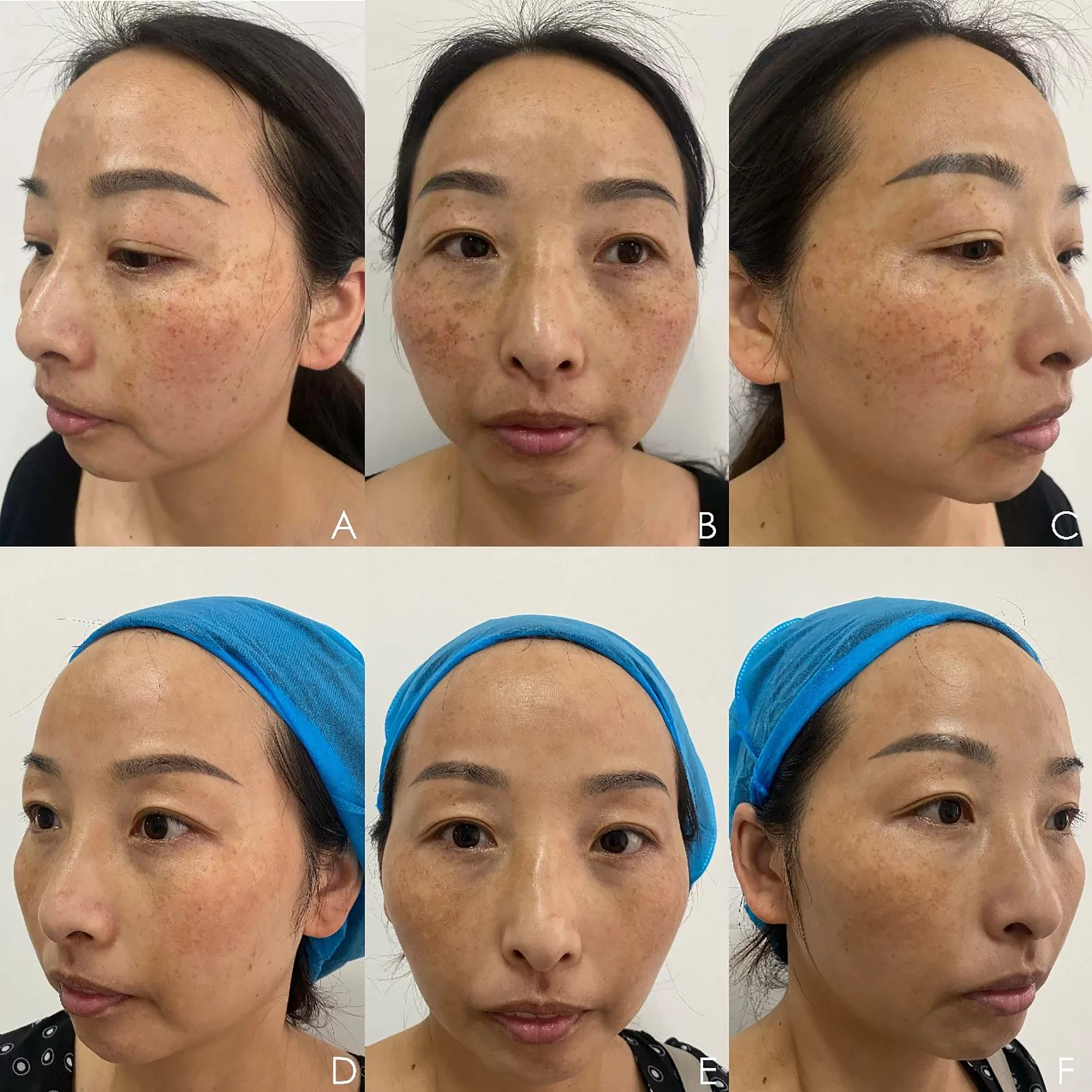
Figure 4. Clinical images from another Group A case, showing pretreatment (A–C) and 2-month posttreatment (D–F) outcomes. Pigmented lesions demonstrated substantial fading, with a more uniform skin tone and diminished contrast against surrounding skin.No serious adverse events occurred in either group. Reported side effects were limited to transient erythema, mild swelling, and occasional bruising, which resolved within several days. No cases of post-inflammatory hyperpigmentation, hypopigmentation, or systemic complications related to oral TA were observed.
Melasma is a chronic, acquired disorder of melanogenesis characterized by localized hypermelanosis on sun-exposed areas, predominantly in adult women. [ 10 ] Its etiology is multifactorial, with major contributors including UV and visible light exposure, hormonal changes such as pregnancy and oral contraceptive use, psychological stress, and genetic predisposition. [ 11 ] Despite multiple therapeutic options, the absence of a gold-standard treatment and the high rate of recurrence continue to pose clinical challenges. [ 12 ]
Conventional therapies include topical agents such as TA and vitamin C, chemical peels, and combinations of QSNY laser with topical or systemic drugs. However, these often require prolonged use and can provoke adverse reactions such as skin irritation or post-inflammatory hyperpigmentation. [ 13 ] Laser-based strategies have therefore become attractive alternatives. The 1064-nm QSNY laser remains the most widely applied modality, but its use at high fluence has been linked to guttate hypopigmentation, rebound melasma, and post-inflammatory pigmentation. [ 14 ] To mitigate these risks, LQSNY toning, based on subcellular selective photothermolysis, has been developed. LQSNY employs a lower fluence (< 3 J/cm 2 ), multiple passes, and larger spot sizes, thereby minimizing inflammation and adverse effects. Mechanistic studies show that LQSNY reduces melanosome numbers, downregulates melanogenesis-associated proteins (tyrosinase-related protein-1 and tyrosinase-related protein-2), and decreases melanogenic stimulators such as α-melanocyte stimulating hormone and nerve growth factor. [ 15 ] It also induces dermal collagen remodeling, leading to improved skin brightness and texture. Although melanin absorbs less energy at 1064 nm compared with shorter wavelengths, the deeper dermal penetration without epidermal injury makes LQSNY especially effective for mixed and dermal-type melasma. In the present trial, however, enrollment was deliberately restricted to the epidermal subtype to ensure a homogeneous study population; extension of these findings to dermal- and mixed-type melasma warrants dedicated investigation. A meta-analysis confirmed significant reductions in MASI and related indices following LQSNY therapy. [ 16 ]
GSH, initially observed serendipitously as a skin-lightening agent, has since been studied extensively. Its mechanisms include direct and indirect inhibition of tyrosinase, shifting melanin synthesis from eumelanin toward pheomelanin, and scavenging reactive oxygen species. It may also modulate the depigmenting effects of other melanocytotoxic agents. [ 17 ] GSH can be delivered topically, orally, or by injection; additional methods include chemical peels, microneedling-assisted application, and mesotherapy. Topical preparations such as creams and lotions are simple and widely available, but intradermal administration ensures higher local bioavailability.
TA, a synthetic lysine analogue and plasmin inhibitor, also shows promise in melasma management. TA blocks the conversion of plasminogen to plasmin, thereby suppressing phospholipase A 2 activation and downstream arachidonic acid–derived mediators such as prostaglandin E 2 and leukotrienes, which stimulate melanogenesis. [ 18 ] In addition, TA shares structural similarity with tyrosine, allowing competitive inhibition of tyrosinase. [ 19 ] By limiting plasmin activity, TA also reduces the release of basic fibroblast growth factor and prevents activation of vascular endothelial growth factor, both potent melanocyte and angiogenic stimulants. [ 20 ] These mechanisms explain its efficacy in UV-induced pigmentation and hormonally driven cases, such as those associated with pregnancy or oral contraceptives.
In this randomized clinical trial, the addition of an intradermal HA-GSH to a regimen of LQSNY laser and oral TA produced earlier and more pronounced improvements in melasma compared with the standard combination of laser and TA alone. The advantage of HA-GSH was evident by the second month of treatment, maintained through the treatment course, and persisted at 2-month follow-up. Patient satisfaction was numerically higher in the combination group, although this difference did not reach statistical significance, and both regimens were well tolerated, with no serious adverse events or pigmentary complications observed.
The trajectory of modified MASI scores supports a clinically meaningful adjunctive effect of HA-GSH. In the baseline-adjusted mixed-effects model, the advantage of HA-GSH was already measurable at the first posttreatment assessment and then remained essentially constant in magnitude across the treatment course, with a nonsignificant group × visit interaction. The benefit is therefore best characterized as an early-onset effect that was subsequently maintained rather than 1 that widened progressively, and the unadjusted group × time interaction did not reach significance once the marked departure from sphericity was taken into account. The persistence of the between-group difference 2 months after the final session nonetheless points to improved short-term durability. Given the chronic and relapsing nature of melasma, these findings highlight the potential of HA-GSH as a valuable adjunct in multimodal treatment strategies. Because melasma is chronic and relapsing, the realistic aim of treatment is durable control and improved quality of life rather than cure. From this perspective, a regimen that achieves earlier and more sustained improvement may reduce the cumulative number of treatment sessions and prolong relapse-free intervals, potentially offsetting its higher per-session cost; moreover, the incremental cost here is limited to a skin-booster add-on layered onto an otherwise standard protocol, and the numerically higher patient satisfaction observed in the combination group, although not statistically significant, is consistent with its perceived value and may support long-term adherence. Nevertheless, we did not perform a formal cost-effectiveness analysis, and the durability advantage was assessed over only a short follow-up; longer studies incorporating relapse rates, the number of maintenance sessions, quality-of-life measures, and health-economic endpoints are needed to determine whether the added cost of HA-GSH is justified in routine practice.
Our findings align with and extend prior evidence supporting multimodal approaches in the treatment of melasma. Several clinical trials have demonstrated that combining oral TA with LQSNY 1064-nm laser achieves superior outcomes compared with either modality alone. For example, a study involving 48 patients showed that oral TA for 8 weeks in combination with 2 sessions of LQSNY laser led to significantly greater mMASI reduction than laser monotherapy, with more patients achieving marked improvement and no serious adverse events. [ 21 ] Similarly, a randomized trial of 60 patients confirmed that adding laser to oral TA provided greater improvement in pigmentation and vascular features on dermoscopy, reinforcing the synergistic potential of targeting multiple pathogenic pathways concurrently. [ 11 ] Beyond systemic adjuvants, topical and injectable agents have also been explored as enhancers of laser efficacy. In a split-face trial of 25 patients, the combination of LQSNY laser with a topical mixture of TA, niacinamide, and kojic acid resulted in significantly greater hemi-MASI improvement than laser alone, highlighting the role of antioxidants and tyrosinase inhibitors in potentiating laser-based pigment clearance. [ 7 ] More recently, injectable regimens incorporating GSH have gained attention. A prospective split-face study of 20 patients demonstrated that intradermal injections of TA, vitamin C, and GSH significantly improved pigmentation parameters with excellent tolerability, underscoring the clinical feasibility of localized antioxidant delivery. [ 3 ] The synergistic role of HA as both a carrier and a dermal modulator has also been suggested. A pilot study of 6 cases found that after laser toning responses plateaued, subsequent injection of non-crosslinked HA enriched with antioxidants yielded further pigment clearance, suggesting that such formulations can overcome treatment resistance and prolong the efficacy of laser therapy. [ 22 ] Similarly, a trial of 29 patients comparing microneedling alone versus microneedling combined with topical GSH showed greater and earlier reductions in mMASI on the GSH-treated side, reinforcing the concept that GSH accelerates and amplifies the effects of procedural interventions. [ 8 ] Taken together, these studies converge on the principle that layering complementary therapies (whether systemic, topical, or injectable) enhances melasma outcomes by simultaneously addressing oxidative stress, tyrosinase activity, melanogenesis, and skin barrier function. Our results build on this foundation by demonstrating that intradermal HA-GSH, when integrated with oral TA and LQSNY laser, provides earlier, greater, and more durable improvements than the established laser–tranexamic regimen alone.
The mechanistic rationale for the observed benefit likely lies in the complementary actions of the 3 components. GSH reduces tyrosinase activity, shifts melanogenesis toward pheomelanin production, and scavenges reactive oxygen species generated by UV exposure. HA serves not only as a carrier but also improves dermal hydration and tissue homeostasis, potentially attenuating post-laser inflammation and stabilizing epidermal turnover. Together with the pigment-targeting action of the laser and the plasminogen-inhibiting effect of TA, this multimodal approach addresses multiple pathogenic pathways in melasma.
Safety outcomes in this trial were favorable. Both regimens were associated only with transient erythema, edema, and mild bruising, which resolved within days. No cases of post-inflammatory hyperpigmentation, hypopigmentation, or systemic complications related to TA were observed. These findings underscore a favorable risk–benefit profile for the combination, particularly in Fitzpatrick skin types III and IV, where the risk of dyspigmentation can be higher.
This study has several limitations. It was conducted at a single center with a relatively small cohort, and the follow-up duration was short, which restricts conclusions regarding long-term durability. Although observer bias was reduced by independent, blinded rescoring of standardized photographs, objective instrument-based pigment quantification (e.g., colorimetry, reflectance spectrophotometry, or image analysis) was not employed, and the inherently subjective nature of visual mMASI scoring remains a limitation. In addition, 14 of the 50 randomized participants (28%) withdrew before completing the protocol. Attrition was balanced between the groups and arose from logistical rather than clinical reasons, but no post-baseline assessments were available for these participants, so the efficacy analysis was necessarily confined to completers and rests on a missing-completely-at-random assumption. Pattern-mixture and tipping-point analyses indicate that the principal finding is robust unless the participants who withdrew from group A would have fared appreciably worse than the group B completers, but residual attrition bias cannot be excluded. The completed sample also fell short of the 50 originally planned; the study was therefore adequately powered for the large between-group difference actually observed but not for smaller effects, and it was clearly underpowered for patient-reported satisfaction, for which no significant difference was demonstrated. Finally, mMASI was recorded to the nearest whole point, which limits the resolution of the instrument, and the unadjusted group × time interaction did not reach significance once the departure from sphericity was taken into account; the conclusion of an adjunctive benefit therefore rests principally on the baseline-adjusted between-group comparisons rather than on a demonstrated difference in the shape of the response trajectories.
Future research should validate these findings in larger, multicenter, assessor-blinded trials with extended follow-up to evaluate relapse rates. Studies optimizing dosing, injection volume, and session intervals are warranted. In addition, incorporation of objective pigment metrics, patient-reported quality-of-life measures, and mechanistic biomarkers such as oxidative stress indices or tyrosinase activity would provide a more comprehensive understanding of treatment efficacy and durability.
The addition of intradermal HA-GSH to LQSNY 1064-nm laser and oral TA provided earlier, greater, and more sustained improvements in melasma with good safety and numerically higher patient satisfaction compared with the standard regimen, although the difference in satisfaction did not reach statistical significance. These findings support HA-GSH as a promising adjuvant in multimodal therapy, although confirmation in larger, long-term studies is needed.
The author would like to express sincere gratitude to her mother, Jianxia Xu, for her unwavering support, encouragement, and understanding throughout the completion of this study and the preparation of this manuscript. Her patience, confidence, and constant encouragement provided invaluable motivation during this work. The author is deeply grateful for her enduring support and inspiration.
Conceptualization: Jie Ling Bai.
Data curation: Jie Ling Bai.
Formal analysis: Jie Ling Bai.
Writing – original draft: Jie Ling Bai.
Writing – review & editing: Jie Ling Bai.
声明:中文精读 · 仅供学术参考。内容来自公开文献检索,不代表本人观点,不构成诊疗建议。 医疗美容需在正规医疗机构由执业医师实施。
基于相同主题推荐 · 继续深挖
随机 1:1 纳入 50 例中国女性表皮型黄褐斑(Fitzpatrick III–IV):A 组为低能量 1064nm Nd:YAG 激光 + 口服氨甲环酸 250mg/日 + 皮
研究类型:单中心回顾性病历回顾研究(非随机、单臂、无同期对照),评估皮秒激光联合透明质酸复合溶液治疗黄褐斑。
提出「HA 填充术后复发性眼睑水肿(PHAREE)」概念,并给出以序贯透明质酸酶 + 分段射频微针为主、必要时辅以 CO2 激光的处置流程。