眶周动态皱纹对下睑成形术手术结果的影响
2026年10月6日24 min read
Plastic and Reconstructive Surgery Global Open
结论与实践要点
预后指标: 术前动态皱纹分级应作为下睑成形术的预后参考。重度动态皱纹患者术后下睑轮廓的“回退”更快(术后1个月至1年间老化征象进展更明显),这并非初始矫正不足,而与眼轮匝肌兴奋性偏高、皮下胶原易损及皮肤偏薄有关;两组在术后1个月早期基线相当,使后续差异更可能归因于皱纹表型本身。
操作建议: 对重度组应更稳健地重建与加固下睑支撑结构(眶隔、眼轮匝肌支持韧带);可考虑术中或分期联合脂肪移植填充泪沟与睑颊沟、肉毒毒素抑制过度肌活动、点阵激光或射频微针刺激胶原,以延缓皱纹再现。但本研究未对术后动态皱纹做序贯标准化再评估,上述辅助手段的叠加价值仍需前瞻性研究验证。
患者沟通: 需向重度动态皱纹患者充分告知“轮廓改善可达成、但老化征象可能更快再现”的预期,以优化满意度;并发症风险不受皱纹严重程度影响,无需因此改变安全预案与围手术期管理。
Background:Lower eyelid bags and periorbital dynamic rhytides are common manifestations of periocular aging in middle-aged and older adult patients seeking aesthetic improvement. Although lower eyelid blepharoplasty corrects bag deformity, its interplay with preoperative dynamic rhytides remains understudied. This retrospective cohort study compares surgical outcomes following lower eyelid blepharoplasty in patients with varying severities of preoperative periorbital dynamic rhytides and to assess differences in treatment efficacy.
Methods:This retrospective study analyzed 56 patients (112 eyes) undergoing transcutaneous lower eyelid blepharoplasty. Patients were stratified into group A (n = 32, severe dynamic rhytides) and group B (n = 24, mild dynamic rhytides) based on the preoperative Lemperle Wrinkle Scale. Postoperative outcomes were evaluated at 1-month and 1-year intervals using Barton and Hirmand grading systems to quantify infraorbital rejuvenation. Secondary endpoints included complication rates and patient satisfaction.
Results:At 1-year follow-up, group A (severe rhytides) showed significantly less improvement in infraorbital contour than group B (mild rhytides) based on standardized grading systems ( P < 0.05). Patient satisfaction was also lower in group A ( P < 0.05). Complication rates did not differ between the groups ( P > 0.05).
Conclusions:The severity of preoperative periorbital dynamic rhytides is associated with less favorable aesthetic outcomes in infraorbital contour after lower eyelid blepharoplasty and predicts lower patient satisfaction. However, it does not increase surgical risks. Preoperative assessment of wrinkle severity should be integrated into surgical planning and patient counseling to optimize expectations.
Question: Does the severity of preoperative periorbital dynamic rhytides predict the rate of infraorbital aging progression after lower eyelid blepharoplasty?
Findings: In this retrospective cohort study of 56 patients (112 eyes), those with severe preoperative dynamic rhytides showed significantly greater progression of infraorbital aging signs between 1 month and 1 year postoperatively, along with lower patient satisfaction, and complication rates were comparable between groups.
Meaning: Severe preoperative periorbital dynamic rhytides predict faster recurrence of infraorbital contour deformity after lower eyelid blepharoplasty and lower patient satisfaction, without increasing surgical risks; preoperative wrinkle assessment should inform surgical planning and patient counseling.
Lower eyelid bags (also termed palpebral bags) represent a prevalent age-related periorbital concern, characterized primarily by anterior herniation of orbital septal adipose tissue due to attenuation of the lower eyelid–retaining structures, often accompanied by tear trough deformities and skin laxity. 1 The pathogenesis is multifactorial, involving involutional changes in the orbital septum, weakening of the orbicularis oculi muscle, and loss of midface support. 2 , 3 Transcutaneous lower blepharoplasty remains a cornerstone procedure for the morphological correction of these contour abnormalities.
Concurrently, periorbital dynamic rhytides—such as crow’s feet, infraorbital wrinkles, and glabellar lines—are another hallmark of facial aging. These are induced by repetitive contractions of the underlying mimetic musculature and, over time, coupled with intrinsic dermal changes, may evolve into persistent static wrinkles. 4 These 2 entities frequently coexist in the aging periorbital region.
Current surgical strategies for lower blepharoplasty are highly effective in addressing fat pseudoherniation and mild to moderate skin excess, with satisfactory early results widely reported. However, the long-term maintenance of these outcomes may be influenced by persistent factors inherent to the patient’s aging phenotype. Periorbital dynamic rhytides—representing both repetitive muscular activity and underlying dermal compromise—are hypothesized to accelerate postoperative reemergence of infraorbital aging signs, even after successful initial contour correction. 5 The impact of preoperative dynamic wrinkle severity on the rate of postoperative aging progression remains unquantified.
Therefore, this study investigates the influence of preoperative periorbital dynamic rhytide severity on the dynamics of postoperative infraorbital aging progression following standard transcutaneous lower eyelid blepharoplasty. We specifically assessed changes in aging signs between 1-month (after edema resolution) and 1-year follow-up. We hypothesized that patients with more severe preoperative dynamic rhytides would exhibit a faster rate of recurrence or progression of infraorbital aging signs during this interval, independent of the initial surgical outcome.
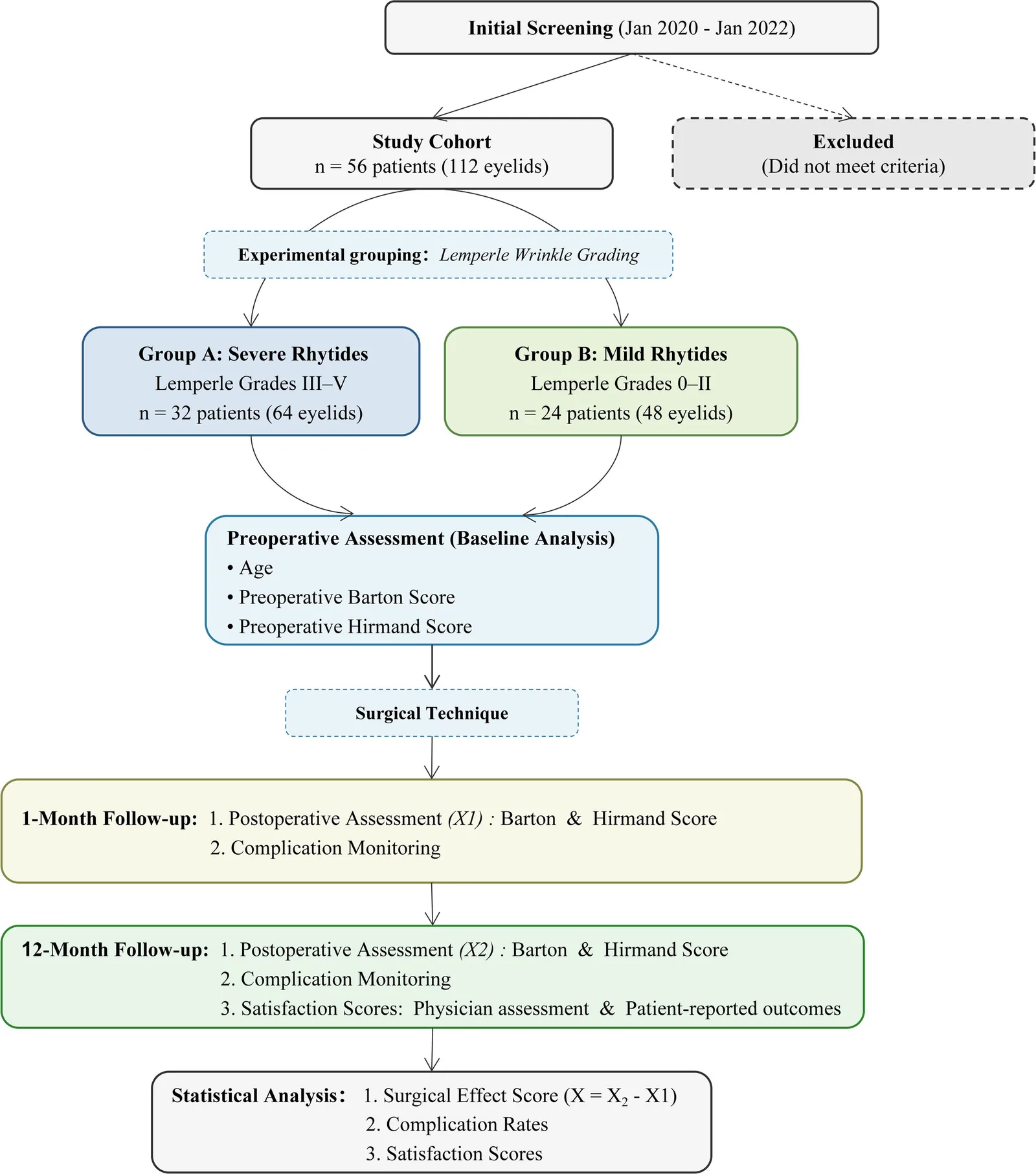
The overall study design is schematically presented in Figure 1 . We conducted a retrospective cohort analysis of 56 consecutive patients (112 eyelids) undergoing primary transcutaneous lower blepharoplasty at our institution (January 2020–January 2022). The cohort comprised 49 female (98 eyelids) and 7 male (14 eyelids) patients, aged 40–63 years (mean 53 ± 6.2 y).
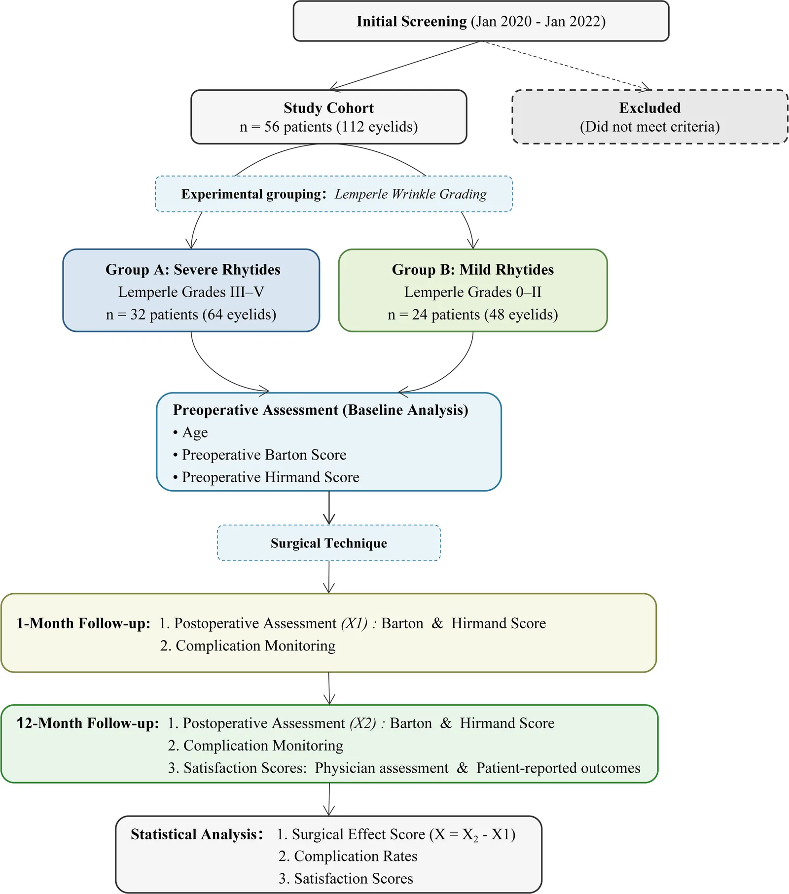
Fig. 1. Study flowchart illustrating patient selection and preoperative wrinkle severity grading (Lemperle scale) to assign groups A and B, surgical intervention, and postoperative assessments at 1 and 12 month(s), with the primary outcome. The surgical effect score (X = X2 − X1) is calculated using the Barton/Hirmand scores to specifically reflect the progression rate of infraorbital aging signs between the 2 follow-up time points.
The institutional review board approved this study, and all participants provided written informed consent.
Periorbital dynamic wrinkles mainly included lateral canthal dynamic wrinkles (crow’s feet), lower eyelid dynamic wrinkles, and glabellar lines. Lemperle et al 6 divided wrinkles into 6 levels:
Before surgery, each patient was asked to perform a maximum voluntary contraction of the orbicularis oculi muscle (eg, by forcefully squinting or executing an exaggerated smile) to evaluate dynamic wrinkle severity. Patients were dichotomized into:
Before comparative outcome analysis, baseline characteristics, including age and preoperative Barton and Hirmand scores, were compared between group A and group B to assess the equivalence of the cohorts at study entry. Preoperative photographs of the patients with facial muscles in the contracted state and the relaxed state were taken from standardized anteroposterior, oblique, and lateral views.
All procedures were performed by senior oculoplastic surgeons using a standardized approach.
Preoperatively, patients were positioned upright to identify and mark orbital fat protrusions. Following standard antiseptic preparation and sterile draping, a subciliary incision was demarcated 1–1.5 mm inferior to the lash line, extending from approximately 2 mm lateral to the lacrimal punctum to 5–10 mm beyond the lateral canthus. Local anesthesia was achieved using a solution containing 5 mL of 2% lidocaine hydrochloride, 5 mL of 0.9% normal saline, and 0.2 mL of epinephrine (1:200,000 concentration), which was infiltrated into the subcutaneous plane of both lower eyelids and infraorbital regions.
Upon achieving adequate anesthesia, a No. 15 scalpel was used to create the skin incision, with careful dissection proceeding to the inferior tarsal border to expose the orbicularis oculi muscle. In cases of muscle laxity, approximately 2–3 mm of redundant orbicularis fibers were resected, with hemostasis achieved using bipolar electrocautery.
The orbital septum was then exposed, and a central septotomy measuring 5–10 mm was performed. Gentle globe pressure facilitated visualization of herniated orbital fat pads (Fig. 2 ). Herniated fat compartments were isolated and excised, and the remaining pedicles were cauterized before release. Residual fat was meticulously repositioned posterior to the orbital septum to prevent postoperative hollowing. This technique was systematically repeated for the medial, central, and lateral fat pads.
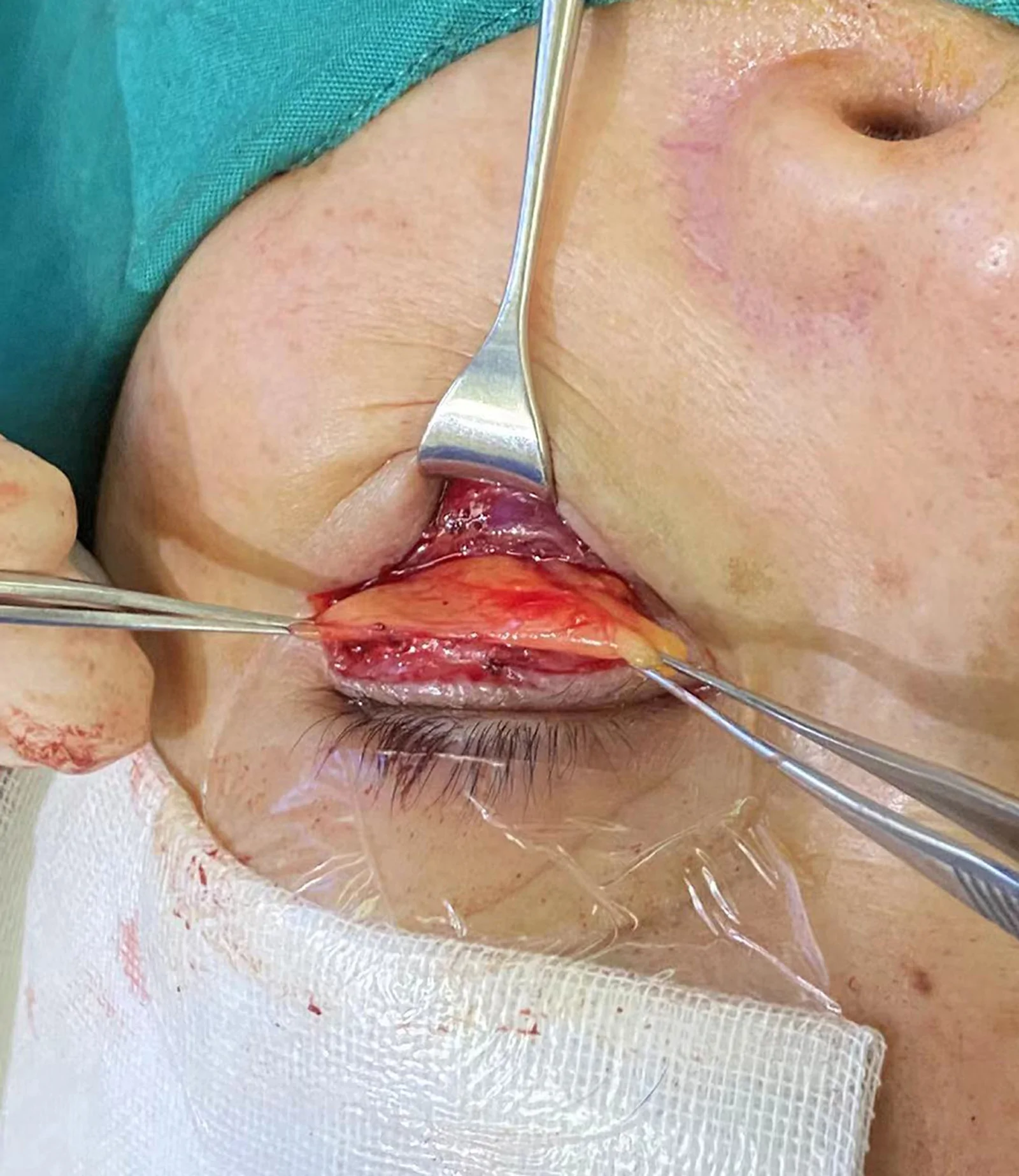
Fig. 2. Intraoperative view of orbital fat management. After exposing the orbital septum and creating a central septotomy of 5–10 mm, gentle globe pressure causes the orbital fat to herniate into view. Excess fat is clamped and excised, whereas the remaining fat is repositioned posterior to the septum to prevent postoperative lower eyelid depression.For patients presenting with tear trough deformities, the medial fat pad was carefully released and advanced inferomedially to fill the preoperative depression. The septal–fat composite flap was then secured to the medial infraorbital periosteum using 6-0 absorbable sutures, followed by layered closure.
Skin excision was guided by having patients open their mouths while directing their gaze superiorly toward the surgeon’s forehead. Redundant skin was gently elevated using Bishop forceps, with the excision line carefully marked to ensure tension-free closure. Hemostasis was verified before wound approximation with 7-0 polypropylene sutures, with meticulous attention to eliminating dog-ear deformities.
The surgical site was dressed with erythromycin ophthalmic ointment and sterile gauze, followed by a cold compress for 30–60 minutes. Sutures were removed on postoperative day 7.
During the 12-month follow-up period, postoperative outcomes were assessed by a panel comprising the operating plastic surgeon and 2 independent plastic surgeons who were blinded to the surgical procedures.
Grades 0, I, II, and III in the 2 grading systems were assigned values of 0, 1, 2, and 3 points, respectively, with higher grades indicating more obvious aging of the inferior orbital region.
To evaluate the postoperative progression of infraorbital aging, we calculated a surgical effect score ( X ). This score was defined as the difference between the Barton/Hirmand score at the 1-year follow-up ( X 2 ) and that at the 1-month follow-up ( X 1 ): X = X 2 − X 1 .
At 1 month postoperatively, the initial contour-correcting effect was evident, and acute edema had largely resolved. Therefore, the change in scores from this time point to 1 year later ( X ) primarily reflects the rate of reappearance or progression of infraorbital aging signs, rather than the magnitude of the initial surgical correction. We acknowledge that residual edema may still have been present at the 1-month postoperative time point, as evident in our representative cases (Figs. 3 , 4 ), and may have partially obscured the final contour, leading to a slight overestimation of the initial surgical effect. However, this potential bias would likely affect both groups equally, and the 1-year endpoint provides a robust assessment after complete remodeling and edema resolution. Therefore, the differential progression rate observed between groups remains valid. This metric allowed us to specifically investigate whether different severities of preoperative dynamic rhytides were associated with distinct long-term aging trajectories after receiving the same initial surgical intervention. Representative pre- and postoperative patient photographs are shown in Figures 3 and 4 .
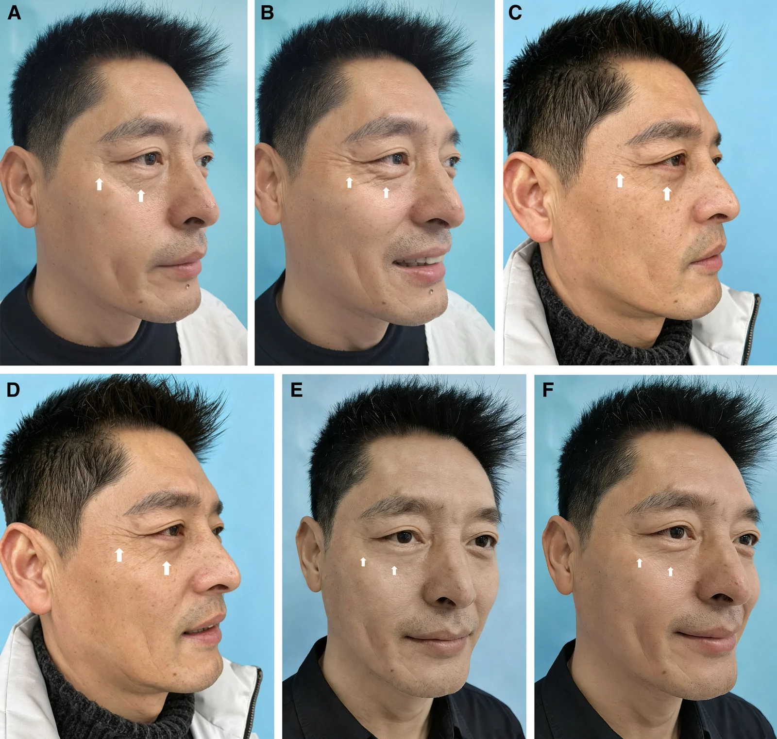
Fig. 3. Representative pre- and postoperative patient photographs, where each patient was asked to perform a maximum voluntary contraction of the orbicularis oculi muscle (eg, by forcefully squinting or executing an exaggerated smile) to evaluate dynamic wrinkle severity (as indicated by the arrows). A 45-year-old male patient (preoperative Lemperle classification was grade V and was classified into group A) is shown preoperatively (A, muscle relaxation; B, muscle contraction), 1 month postoperatively (C, muscle relaxation; D, muscle contraction), and 1 year postoperatively (E, muscle relaxation; F, muscle contraction), demonstrating the surgical effects of lower eyelid bag removal. Note the presence of mild residual edema at the 1-month postoperative view, which typically resolves by 3–6 months.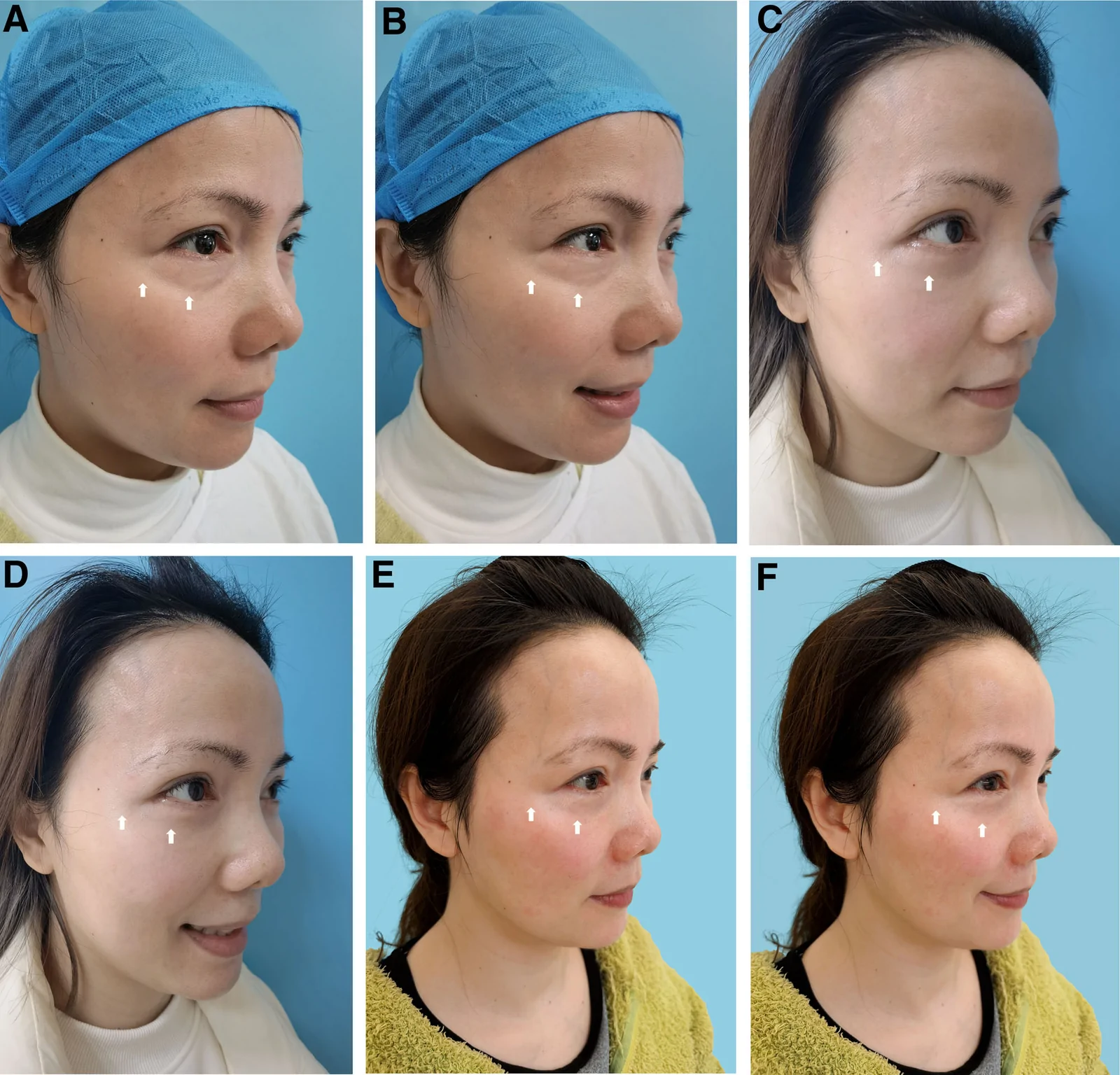
Fig. 4. Representative pre- and postoperative patient photographs in which each patient was asked to perform maximum voluntary contraction of the orbicularis oculi muscle (eg, by forcefully squinting or executing an exaggerated smile) to evaluate dynamic wrinkle severity (as indicated by the arrows). A 43-year-old female patient (preoperative Lemperle classification was grade II and was classified into group B) is shown preoperatively (A, muscle relaxation; B, muscle contraction), 1 month postoperatively (C, muscle relaxation; D, muscle contraction), and 1 year postoperatively (E, muscle relaxation; F, muscle contraction), demonstrating the surgical effects of lower eyelid bag removal. Note the presence of mild residual edema at the 1-month postoperative view, which typically resolves by 3–6 months.
Documented complications during the 12-month follow-up period included incision swelling, infection, scar hyperplasia, subcutaneous ecchymosis, hematoma, lower eyelid retraction, eversion, numbness, foreign body sensation, dry eye, retrobulbar hematoma, decreased vision, and eyelash loss. The postoperative complication rate (%) was calculated as the number of eyes with complications divided by the total number of eyes multiplied by 100.
All evaluators used a standardized Likert scale to objectively grade the therapeutic outcomes bilaterally. Concurrently, patient-reported outcome measures were collected to evaluate overall satisfaction with the surgical intervention.
Continuous variables were analyzed using independent t tests, whereas categorical data were compared using χ 2 tests (SPSS, version 26.0; α = 0.05). The data for each group were graphed using GraphPad Prism 10.0.2.
Comparative analysis of baseline demographics and clinical characteristics confirmed that the 2 groups were well balanced at study entry. ( See table, Supplemental Digital Content 1 , which compares baseline characteristics [age, sex, preoperative Barton and Hirmand scores], postoperative surgical effect scores [ X = 1‑year score − 1‑month score for both grading systems], and complication rates between group A [severe rhytides] and group B [mild rhytides], https://links.lww.com/PRSGO/F255.) No statistically significant differences were observed between group A and group B in mean age (52.75 ± 1.23 versus 54.08 ± 1.39 y; P = 0.421), preoperative Barton score (2.66 ± 0.06 versus 2.63 ± 0.07; P = 0.734), or preoperative Hirmand score (2.19 ± 0.10 versus 2.13 ± 0.10; P = 0.642).
We conducted comparative analyses of infraorbital aging progression using standardized grading systems: group A demonstrated significantly greater aging progression at the 1-year follow-up ( Supplemental Digital Content 1 , https://links.lww.com/PRSGO/F255).
There was no significant difference between the 2 groups ( Supplemental Digital Content 1 , https://links.lww.com/PRSGO/F255). The postoperative complication rate (%) in group A was 15.6% (10 of 64) and that in group B was 14.6% (7 of 48) (χ 2 = 0.023; P = 0.879 > 0.05).
The observed complications were primarily transient. Transient ecchymosis (n = 5) resolved spontaneously within 1–2 weeks. Dry eye (n = 8) occurred and was effectively managed with sodium hyaluronate eye drops. More serious complications, such as incision infection and lower eyelid retraction, did not occur.
Significantly higher satisfaction rates were observed in group B. ( See table, Supplemental Digital Content 2 , which presents surgeon‑assessed satisfaction with surgical outcomes [per eye] and patient‑reported postoperative satisfaction [per case], both stratified by group and expressed as the percentage of cases rated as “satisfied” [very satisfactory + satisfactory] cases, https://links.lww.com/PRSGO/F256.)
According to the collected evaluations of doctors’ satisfaction with the surgical results, the rate of satisfaction with the surgical results was 91.7% (44 of 48) in group B and 75.0% (48 of 64) in group A (χ 2 = 4.120; P = 0.042 < 0.05) ( Supplemental Digital Content 2 , https://links.lww.com/PRSGO/F256).
Regarding patient satisfaction after surgery, 91.7% (22 of 24) of patients in group B were satisfied with the postoperative effects compared with only 65.6% (21 of 32) of patients in group A (χ 2 = 3.159; P = 0.049 < 0.05) ( Supplemental Digital Content 2 , https://links.lww.com/PRSGO/F256).
Lower eyelid bags are more common in middle-aged and older adult people. Historically, researchers attributed the formation of bags to skin laxity and increased orbital fat, advocating maximal fat and skin removal. However, recent observations have indicated that lower eyelid bags result from an imbalance between orbital fat mass and the supporting structures, 9 – 11 rather than merely an increase in fat volume. With age, the supporting structures of the lower eyelid weaken, which includes laxity of the skin and orbital septum, elongation of the orbicularis oculi muscle and its supporting ligament, and weakening of the retraction strength. 12 Additionally, midface deep fat compartment atrophy variably occurs, weakening upper tissue support and causing downward movement of superficial fat pads 13 , 14 ; orbital bone absorption and retraction 15 , 16 contribute to the high incidence of lower eyelid bags in middle-aged and older adult patients. Intraoperatively, we observed that some patients had minimally increased orbital fat volume, but due to weakened supporting structures, even a normal volume of orbital fat could protrude anteriorly, causing lower eyelid bags. 13
Periorbital dynamic rhytides occur mainly when the facial expression muscles contract. Contraction of the muscle fibers of the orbicularis oculi, corrugator supercilii, and depressor supercilii transmits movement to the upper layer of skin through fibrous tissue attachments, resulting in the formation of folds. In young individuals, skin resilience and subcutaneous toughness allow rapid recovery upon muscle relaxation, making rhytides less apparent. In contrast, aging skin undergoes irreversible collagen breakdown, reducing periorbital skin resilience and preventing rapid recovery. Due to differences in race, age, skin aging, and muscle contraction strength, periorbital dynamic rhytides exhibit varying manifestations and grades. 17 , 18 The facial wrinkle grading method proposed by Lemperle et al 6 remains one of the most commonly used methods.
To ensure that observed differences could be attributed to rhytide severity rather than confounding baseline characteristics, we compared key preoperative variables between groups. Results confirmed that group A and group B were well matched in mean age and preoperative lower eyelid bag severity (assessed using the Barton and Hirmand scores, all P > 0.05) ( Supplemental Digital Content 1 , https://links.lww.com/PRSGO/F255). This equilibration of baseline demographics and surgical indications strengthens the inference that the subsequent divergence in aging progression was associated with the predefined factor of wrinkle severity.
For surgery in both groups, a lower eyelid margin approach (external incision method) was used. Orbital septal fat was appropriately removed and repositioned, the lower eyelid–supporting structures were reconstructed and reinforced, and the skin was conservatively excised.
Our findings demonstrate that patients with severe preoperative dynamic rhytides (group A) exhibited a significantly greater increase in Barton and Hirmand scores between the 1‑month and 1‑year postoperative assessments compared with those with mild wrinkles (group B). This differential rate of score progression suggests that severe dynamic rhytides are associated with an accelerated pace of infraorbital aging after surgical contour correction. We postulated that this acceleration is not merely a reflection of poorer initial surgical outcomes—as both groups started from a comparable early postoperative baseline at 1 month—but rather stems from 2 interrelated mechanisms inherent to the severe rhytides phenotype.
This may be caused by excessive contraction of the orbicularis oculi muscle. In group A, patients had obvious dynamic rhytides and a low orbicularis oculi excitability threshold; frequent muscle contraction stimulation accelerated the degenerative rupture of subcutaneous collagen fibers. Additionally, the periorbital skin was thinner; there was less, but flaccid, subcutaneous tissue; and aging around the eyes was more likely to appear, thus affecting the surgical effect after lower eyelid bag removal, manifesting as re-relaxation of the lower eyelid structure. However, due to the intraoperative removal of part of the protruding orbital septum fat and the restoration of the basic shape of the orbital septum, 19 the postoperative lower eyelid bag shape improved compared with the preoperative appearance. In group B, although orbicularis oculi contractility was weak and some patients showed muscle laxity, intraoperative reinforcement and reconstruction of the supporting structures prevented short-term re-relaxation, resulting in good postoperative outcomes. Collectively, our findings indicate that severe preoperative dynamic rhytides serve as a significant baseline predictor of a faster rate of infraorbital aging progression and less favorable long-term contour outcomes following lower eyelid blepharoplasty.
Regarding the postoperative satisfaction score, satisfaction with the elimination of obvious periorbital dynamic rhytides was significantly lower in group A than in group B ( P < 0.05).
No significant difference was observed in postoperative complications between the 2 groups. Because the periorbital skin was thin, the subcutaneous tissue was flaccid, and compression was difficult, incomplete hemostasis might have led to hematoma or congestion. Due to sensitive nerve distribution, postoperative dry eye or foreign body sensation was a common complication. 20 For postoperative dry eye, we used sodium hyaluronate eye drops with satisfactory efficacy. These results indicate that dynamic rhytides do not increase surgical risk and that they primarily influence aesthetic perception rather than surgical safety.
Our finding that severe dynamic rhytides predict accelerated aging recurrence suggests that adjunctive perioperative strategies may optimize long-term outcomes. Although botulinum toxin remains the standard treatment for reducing hyperkinetic orbicularis activity, it addresses only the muscular component of wrinkle formation. Given that patients with severe rhytides also exhibit compromised dermal quality and volumetric deficits, a multimodal approach may be beneficial. For instance, fat grafting to the tear trough and palpebromalar grooves can restore volume loss and provide a biological scaffold that may counteract recurrent laxity. 21 Additionally, energy-based modalities such as fractional laser resurfacing 22 or radiofrequency microneedling 23 can stimulate neocollagenesis and improve dermal architecture, potentially slowing wrinkle recurrence. Future prospective studies should investigate whether combining lower blepharoplasty with such procedures (eg, intraoperative fat grafting or staged laser therapy) yields more durable infraorbital rejuvenation in patients with severe dynamic rhytides.
These insights shift clinical focus from merely assessing static preoperative wrinkles to considering their prognostic value for long-term aging dynamics. For patients with severe dynamic rhytides, surgeons might consider (1) more robust reinforcement of lower eyelid support structures; (2) adjunctive procedures targeting hyperkinetic musculature (eg, neuromodulator therapy); and (3) proactive counseling regarding the potential for faster recurrence of aging signs.
This study has several limitations. First, its retrospective design inherently limits control over all confounding variables. Second, although our primary outcome measured the progression of contour-based aging signs, we did not serially reassess dynamic rhytides postoperatively using a standardized scale. Thus, we cannot definitively state whether the wrinkles worsened, improved, or remained unchanged, or how that change directly correlated with contour progression. However, our design specifically aimed to isolate the influence of the preoperative wrinkle phenotype on contour aging dynamics rather than to track wrinkle evolution. Future prospective studies incorporating serial and multimodal assessments may further elucidate the interplay between these aging components.
The severity of preoperative periorbital dynamic rhytides is associated with less favorable aesthetic outcomes in infraorbital contour after lower eyelid blepharoplasty and predicts lower patient satisfaction. However, it does not increase the risk of postoperative complications. Preoperative assessment of wrinkle severity should be integrated into surgical planning and patient counseling to optimize expectations.
The authors have no financial interests or commercial associations to disclose in relation to the content of this article. No conflicts of interest exist for any of the authors.
Given the retrospective design of this study, additional individual informed consent for data analysis was waived; however, all patients provided routine surgical informed consent preoperatively, which included permission for the use of their anonymized images for research publication.
The authors sincerely thank the partners for providing valuable comments on the design and conduct of this study.
This study, involving human participants, was reviewed and approved by the ethics committee of the Medical University of Wenzhou (KY2023-126), Zhejiang, China.
补充视频 / 附件(原文未随文提供原图,可于出版方页面查看)gox-14-e8104-s001.pdf补充视频 / 附件(原文未随文提供原图,可于出版方页面查看)gox-14-e8104-s002.pdf
声明:中文精读 · 仅供学术参考。内容来自公开文献检索,不代表本人观点,不构成诊疗建议。 医疗美容需在正规医疗机构由执业医师实施。
基于相同主题推荐 · 继续深挖
前瞻性单盲随机对照试验,30例按Simon分型分为两组各15例。
一项回顾性队列研究(56例/112眼)按术前 Lemperle 皱纹分级,将经皮肤下睑成形术患者分为重度动态皱纹组(A组,III–V级,n=32)与轻度组(B组,0–II级,n=2
V3中面部提升经睫毛下切口,沿内眦线、外眦线、颧脂肪垫下界三个向量悬吊,可同时改善中面部下垂、下睑袋及睑颊轮廓不连续。