超声引导腹直肌脂肪移植用于腹部雕刻:20例MRI影像评估
2026年10月9日21 min read
Plastic and Reconstructive Surgery Global Open
结论与实践要点
本研究为前瞻性影像评估的病例系列,纳入20例接受超声引导腹直肌脂肪移植(UGRAFT,用于腹部雕刻)的患者(男6/女14,平均37.0岁,平均BMI 25.6),以术前与术后6个月MRI测量腹直肌中段与下段厚度。主要结局:术后6个月肌段总厚度(肌肉+移植脂肪)较术前显著增加(男2.39 vs 1.59 cm、女1.86 vs 1.25 cm、合计2.02 vs 1.36 cm,均P<0.05),提示移植脂肪获得一定留存并改善肌腹轮廓。
局限性:样本量小(n=20)、随访仅6个月、缺乏随机对照与长期(>1年)脂肪存活率数据;脂肪厚度单独测量在术后有所下降,肌腹“增厚”部分来自移植物而非纯肌肉增生。作者提出脂肪移植或具肌肉再生潜能属初步假说,尚待基础研究证实,本文未提供在体机制证据。该技术在健身人群的腹部雕刻中具应用前景,但应如实告知患者脂肪吸收与轮廓回退的可能,避免过度承诺。
Background:Muscle enhancement through autologous fat grafting has become an important aspect of body contouring procedures, targeting several muscle groups, including the rectus abdominis, deltoids, biceps, trapezius, longissimus, hamstrings, and vastus muscles.
Methods:Patients who underwent the ultrasound-guided rectus abdominis fat transfer technique were evaluated using preoperative and postoperative magnetic resonance imaging (MRI) scans to assess the anatomy of the rectus abdominis muscles. Preoperative MRI measured the muscle thickness of the middle and lower rectus abdominis segments on both sides, as they were the most representative. Postoperative imaging was performed 6 months after surgery, assessing the total thickness of the rectus abdominis segments (including fat graft and muscle), the muscle thickness alone, and the remaining fat graft thickness.
Results:The ultrasound-guided rectus abdominis fat transfer technique was performed on 20 patients between September 2022 and April 2023. Of these, 6 were men and 14 were women, with a mean age of 37.05 years (range: 25–57 y). Postoperative MRI revealed a statistically significant increase in the total thickness of the muscle segments (including muscle and fat) compared with preoperative measurements.
Conclusions:Ultrasound-guided fat grafting is a safe and effective procedure currently used in abdominal etching. Long-term MRI assessments showed significant fat retention and rectus abdominis muscle enhancement.
Question: How does the intramuscular fat graft amplification evolve over time?
Findings: The preoperative average total thickness measurement of the muscle packs and the total thickness measurements at 6 months postoperatively show a significant increment (muscle and fat); these differences were statistically significant.
Meaning: Ultrasound-guided fat grafting is a safe and effective procedure used in abdominal etching nowadays. Magnetic resonance imaging in long-term patients showed significant fat retention in association with muscular rectus abdominis enhancement.
Body contouring procedures have become increasingly popular as part of the trend to achieve an athletic figure, with muscle enhancement playing an important role in their success. 1 , 2 Fat grafting has long been used for muscle enhancement, primarily targeting subfascial or subcutaneous areas. It is especially useful in reconstructive surgery for oncological patients or those with deformities. 3 – 6
In recent years, autologous fat grafting for muscle enhancement has emerged as an important part of body contouring procedures. This technique enhances several muscle groups, including the rectus abdominis, external and internal obliques, deltoids, biceps, trapezius, longissimus, hamstrings, and vastus muscles. 7 , 8 – 10
The ultrasound-guided rectus abdominis fat transfer (UGRAFT) technique involves the use of ultrasound to guide fat transfer to the rectus abdominis muscle, combined with abdominal liposuction, to create a more natural abdominal contour. 11 , 9 A common concern regarding intramuscular fat grafting is the potential risk of thrombosis or muscle injury. However, studies show that when performed with comprehensive knowledge of anatomy and adherence to appropriate protocols, these risks can be significantly reduced. 9 , 12
Although muscle enhancement through autologous fat grafting visibly increases muscle volume, its primary effect is on muscle tissue, with muscle regeneration being an important part of the objective of these techniques. Fat grafting introduces not only autologous tissue but stem cells (SCs) as well, which are present in large quantities and have regenerative properties. This promotes long-term enhancement and reduces the resorption that can occur in grafts in other areas. 13 , 14 Although it is not possible to determine how long muscle enhancement through autologous fat grafting will last, using adequate liposuction and fat preparation techniques is known to ensure high-quality grafts, which will help achieve favorable outcomes. 15 – 19 The aim of this study was to assess the retention of fat grafts in the rectus abdominis muscles using the UGRAFT technique and to evaluate its effect on muscle thickness.
A total of 20 patients (6 men and 14 women) seeking body contouring surgery were recruited from the Dr. Mauricio Viaro Clinic in Santa Maria, Rio Grande do Sul, Brazil. All patients were evaluated and operated on by the same board-certified plastic surgeon (Dr. Mauricio Viaro) between September 2022 and April 2023.
Each participant received a detailed explanation of the procedure and the study, and all provided written informed consent. The study complied with the provisions of the Declaration of Helsinki.
Inclusion criteria for the study required patients to seek body contour improvement through body contouring surgery, undergo the UGRAFT procedure as part of their treatment, be between 18 and 60 years old, have a body mass index of less than 30 kg/m 2 , and achieve a surgical score of 2 or lower based on the Goldman cardiac risk index. Patients with a history of massive weight loss, bariatric surgery, prior abdominal surgery, or prior body contouring surgery were excluded.
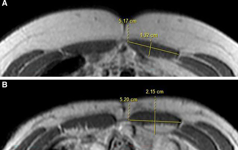
All patients underwent standard preoperative evaluations for body contouring surgery. In addition, preoperative and postoperative magnetic resonance imaging (MRI) was performed for anatomical assessment of the rectus abdominis muscle (Figs. 1 , 2 ). The preoperative assessment focused on measuring the thickness of the middle and lower segments of the rectus abdominis muscle on both sides (right and left inferior “packs,” and right and left middle “packs”), which were the most representative, totaling 80 muscle segments in 20 patients.
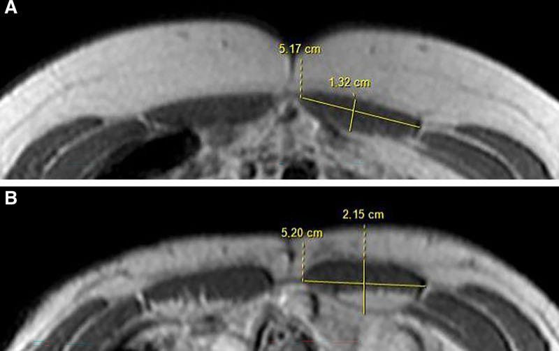
Fig. 1. MRI study of a 43-year-old woman with a 40-cc fat graft in the rectus abdominis muscles. A, Preoperative MRI. B, Postoperative MRI after 6 months.超声引导下腹直肌脂肪移植的手术规划与体表标记示意。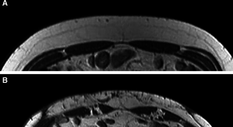
Fig. 2. MRI study of a 30-year-old man with a 60-cc fat graft in the rectus abdominis muscles. A, Preoperative MRI. B, Postoperative MRI after 6 months.Postoperative MRI assessment was performed 6 months after surgery to measure the combined thickness of the muscle and fat graft in the same rectus abdominis segments, as well as muscle thickness alone and the thickness of the remaining graft. Measurements were taken in the anterior–posterior plane at the midpoint of each muscle belly, at an equidistant point from each tendon intersection, before and after UGRAFT (Fig. 3 ).
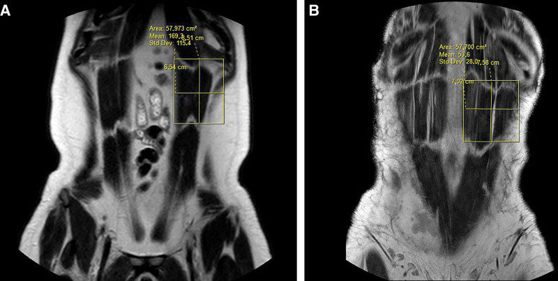
Fig. 3. Measurements taken in the anteroposterior plane at the midpoint of each muscle belly, at a point equidistant from each tendon intersection. A, Before UGRAFT. B, After UGRAFT.Fat volume was calculated as the percentage observed in postoperative images. Two certified radiologists independently reviewed the images, blinded to the patients’ identities and the amount of fat grafted. The UGRAFT technique was performed according to the description of the original technique. 11
The preoperative workup included a series of laboratory tests and imaging studies, including ultrasonography and MRI of the abdominal wall. To optimize graft survival, it is recommended that patients’ nutrition and overall health be maximized before autologous fat transfer. Therefore, we initiated the NutrUgraft supplementation protocol 30 days before surgery, which includes COQ10 (100 mg, 1 capsule in the morning and 1 in the evening), Dimpless (Galena, 40 mg, 1 capsule in the evening), Biointestil (Infinity Pharma, 300 mg, 1 capsule at lunch), IBS Care (Lemma, 100 mg, 1 capsule in the evening), and Impact dietary supplement (Nestlé, 1 unit daily for 7 d). 20
In addition, supplements were prescribed to support muscle regeneration, including vitamin C, glutamine, zinc, quercetin, bromelain, and B-complex vitamins. These micronutrients are clinically proven to play a crucial role in wound healing and enhance graft viability following surgery. 21 , 22
The Monaco-Rios functional protocol was implemented 15 days before surgery. This protocol focuses on metabolic and hormonal optimization through nutritional and caloric restriction, using a ketogenic diet and intermittent fasting associated with 30 minutes of daily physical activity. This approach progressively reduces the patient’s inflammatory process, leading to a better metabolic response to surgical trauma.
Ringer’s lactate solution was administered in the immediate preoperative period, before induction of anesthesia, with subsequent fluid losses managed according to liposuction output and urine output. Deep vein thrombosis prevention measures included the use of pneumatic compression boots and enoxaparin for 10 days postoperatively. Antibiotic prophylaxis with a first-generation cephalosporin (cefazolin) was administered to all patients.
All preoperative markings were performed under ultrasound guidance, allowing for the identification of the degree of diastasis recti and the linea semilunaris, clearly observed at the junction between the lateral border of the rectus abdominis muscles and the lateral abdominal muscles (obliques and transverse). The abdominal “packs” (segments) were also designed based on ultrasound visualization.
For imaging, a linear probe greater than 7.5 MHz connected to an Apple iPad as the display was used. The patients’ rectus abdominis muscles were rescanned while lying on both the right and left sides. Then, each patient’s abdominal “pack” was identified and marked with methylene blue, creating definitive landmarks for fat transfer. ( See Video 1 [online] , which displays 6-month postoperative results with the UGRAFT technique in a 24-y-old woman.)
For muscle access, twin Viaro cannulas (Alice and Sophie, 2.5 and 3.0 mm in diameter and 17 and 25 cm in length, respectively) were used. An incision was made in the inguinal region, using the same access port as for liposuction, just below the linea semilunaris. The cannula, aligned parallel to the linear probe, was inserted into the inferior segment of the rectus abdominis muscle at a 45-degree angle. After loss of resistance was noted, the intramuscular plane was located, and 20-mL boluses were administered at 3 distinct sites (total volume of 60 mL): along the linea semilunaris (at the lateral edge of the inferior rectus), at the upper limit of the inferior tendinous intersection (“first pack”), and at the lower limit of the rectus abdominis attachment to the pubic tubercle. After fat transfer to the lower lateral border, the cannula was advanced beyond the first muscle belly toward the lower segment, and fat graft was distributed across 2 or 3 sites according to patient needs.
After crossing the intersection in a longitudinal ultrasound view, the probe was rotated to a transverse (cross-sectional) view, showing only the tip of the cannula and the lower segment in the transverse direction. The optimal location for fat transfer is subaponeurotic, just beneath the anterior rectus sheath, and the fat should be injected in boluses. An anterior and central 20-mL bolus was initially injected under direct ultrasound visualization. If the fat distribution was deemed ideal (demonstrating optimal fat-muscle interaction and dispersion), an additional 20-mL bolus was injected in the same location. The cannula was then advanced to the lateral region, where another 20-mL bolus was injected, followed by a final 20-mL bolus in the medial region. A total volume of 40–80 mL was administered in the lower segment. ( See Video 2 [online] , which displays the presentation of surgical steps.)
The other segments were approached via the umbilical entry point. The muscle is typically accessed at the level of the lower segment, which had already been grafted in the previous step. After the Viaro cannula entered the lower segment, the ultrasound probe was maintained in a longitudinal view to guide the cannula toward the middle segment, overcoming the resistance of the middle tendinous intersection. After successfully entering the middle segment, the probe was rotated to obtain a cross-sectional view. At this point, a central bolus of 20–40 mL was administered, followed by lateral and medial boluses, each approximately 20 mL in volume. This resulted in a total fat transfer volume of 40–80 mL ( see Video 2 [online] ).
To approach the upper segment, the cannula was maintained in place after grafting the middle segment, and the ultrasound probe was shifted back to a longitudinal view. This allowed direct visualization of the superior tendinous intersection. After passing this intersection, a subaponeurotic bolus of 20–40 mL was administered inside the upper segment. The cannula was then slightly withdrawn, without reentering the middle segment, and repositioned more centrally or laterally within the upper segment. With the probe placed over the lower ribs (showing the cannula and costal margin on the display), a final 20-mL bolus was administered as anteriorly as possible. The upper segment typically receives between 40 and 80 mL of fat, but this volume is generally less than that used in the middle and lower segments due to its smaller capacity ( see Video 2 [online] ). All patients underwent standard preoperative assessment for body contouring surgery and preoperative and postoperative MRI, as previously described (Fig. 2 ) ( see Video 1 [online] ).
补充视频 / 附件(原文未随文提供原图,可于出版方页面查看)gox-13-e6877-s001.mp4Video 1. This video shows the 6-month postoperative results.补充视频 / 附件(原文未随文提供原图,可于出版方页面查看)gox-13-e6877-s002.mp4Video 2. This video shows the presentation of surgical steps.
Statistical analyses were performed using Stata/IC 15.0 (StataCorp, College Station, TX). For quantitative variables, measures of central tendency and dispersion were calculated, whereas categorical variables were expressed in absolute and relative frequencies.
To assess the normality of the data, the Shapiro–Wilk test was performed. For the comparative analysis of rectus abdominis muscle thickness before and 6 months after surgery, a paired t test (Student t test for paired samples) was conducted. A P value of less than 0.05 was considered statistically significant.
Body contouring surgery using the UGRAFT technique was performed on 20 patients between September 2022 and April 2023. Among these patients, 6 were men and 14 were women, with a mean age of 37.05 years (range: 25–57 y) and a mean body mass index of 25.56 kg/m 2 . Patients engaged in physical activity an average of 2.81 times per week (Table 1 ).
Table 1. Descriptive Statistics
| N | Age, Mean, Y | Body Mass Index, kg/m 2 | Exercise Frequency per Week | |
|---|---|---|---|---|
| Men | 6 | 35 ± 3.35 | 26.8 ± 1.11 | 2.5 ± 2.08 |
| Women | 14 | 37.93 ± 11.15 | 25.01 ± 2.64 | 2.92 ± 1.16 |
| Total | 20 | 37.05 ± 9.48 | 25.56 ± 2.38 | 2.81 ± 1.38 |
The mean volume of fat transferred to each muscle segment is shown in Table 2 . Mean rectus abdominis muscle thickness measured preoperatively and at 6 months postoperatively is shown in Table 3 . Both male and female patients demonstrated a statistically significant increase in muscle thickness (Fig. 4 ).
Table 2. Mean Fat Transfer per Muscle Pack of the Rectus Abdominis Muscle
| Rectus Abdominis Pack | Mean Fat Graft, mL |
|---|---|
| Righ intermediate | 47.5 ± 16.09 |
| Left intermediate | 47.25 ± 16.5 |
| Right inferior | 48.75 ± 19.18 |
| Left inferior | 48.75 + 19.18 |
| Total | 48.13 + 17.47 |
Table 3. Average Thickness of Rectus Abdominis Muscle Packs
| Men, cm | Women, cm | Total, cm | |
|---|---|---|---|
| Before surgery | 1.59 ± 0.36 | 1.25 ± 0.21 | 1.36 ± 0.31 |
| After surgery (6 mo) | 2.39 ± 0.39 | 1.86 ± 0.33 | 2.02 ± 0.43 |
| N | 6 | 14 | 20 |
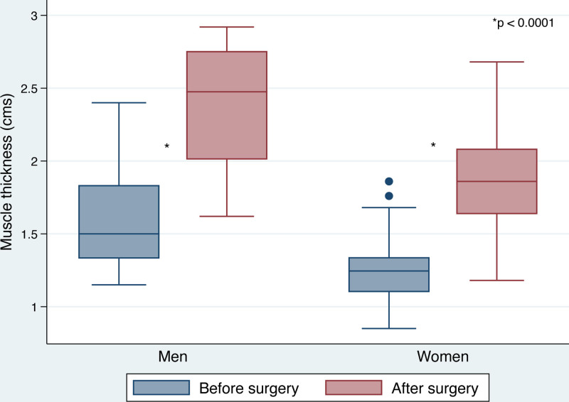
Fig. 4. Box plot showing the mean thickness of the rectus abdominis muscle segments by sex, before and 6 months after surgery.按性别分组的术前与术后6个月腹直肌段厚度箱线图(差异具统计学意义)。For the lower muscle segments, a differentiated analysis of postoperative measurements was conducted, comparing total thickness (including muscle and fat) with muscle thickness alone. Figure 5 illustrates this distinction, showing a reduction in thickness when considering only muscle tissue. However, when compared with preoperative muscle thickness, the difference remains statistically significant (Figs. 6 , 7 ).
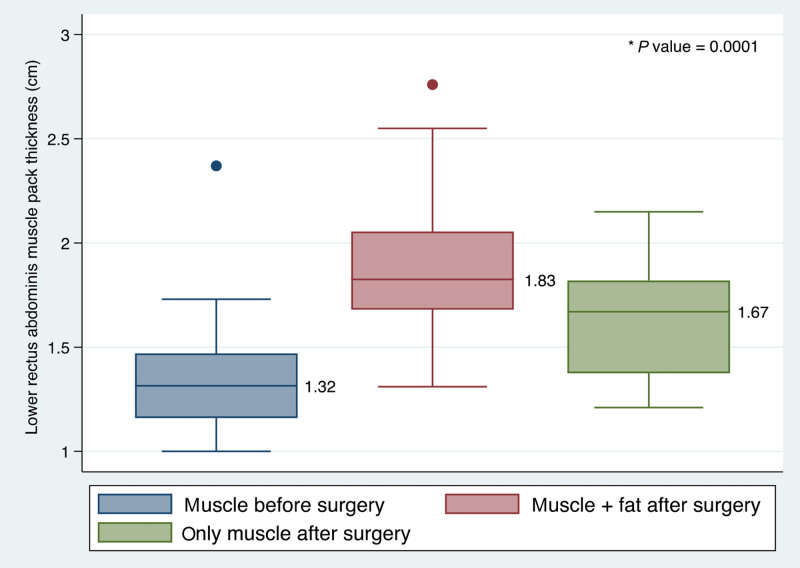
Fig. 5. Box plot showing the differentiated thickness measurement of the lower rectus abdominis muscle segments, before and 6 months after surgery.下腹壁肌段分层厚度对比(含移植脂肪 vs 仅肌肉组织)。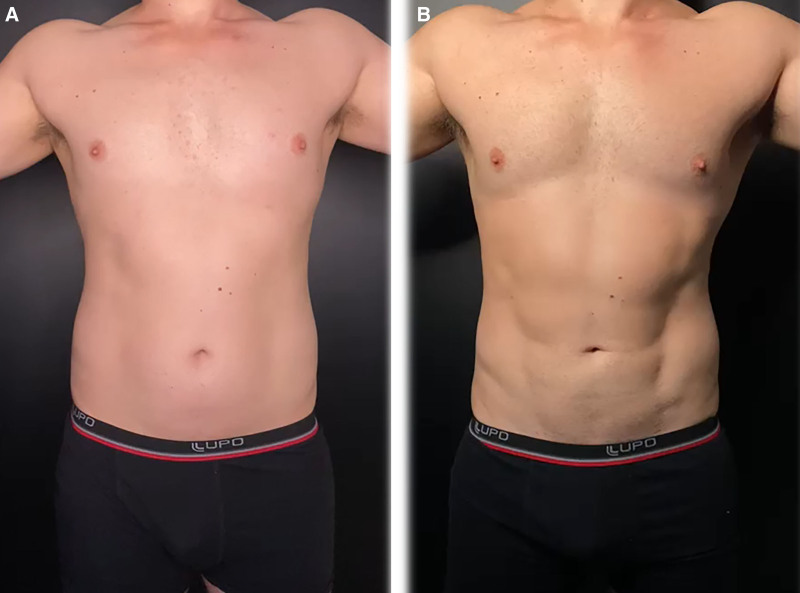
Fig. 6. A 35-year-old male patient. A, Frontal view preoperatively. B, Frontal view 6 months after the UGRAFT technique, with 60-mL fat graft in each rectum abdominis muscle.35岁男性病例:术前与术后6个月正面对照(每侧腹直肌移植约60 mL)。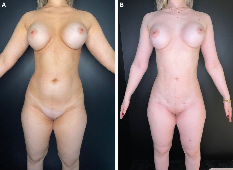
Fig. 7. A 24-year-old female patient. A, Frontal view preoperatively. B, Frontal view 6 months after the UGRAFT technique, with 30-mL fat graft in each rectum abdominis muscle.代表性病例术前与术后6个月对照,展示腹部雕刻轮廓改善。
Skeletal muscles have an inherent ability to regenerate after injury. Regeneration of skeletal muscle is an extremely complex biological process, which involves multiple cell types. Skeletal muscle SCs (also known as satellite cells) are crucial for muscle development, growth, maintenance, and repair. Cell fates and function have been extensively investigated in the context of skeletal muscle regeneration. In addition to SCs, several other cell types—including fibro-adipogenic precursors, endothelial cells, fibroblasts, pericytes, and certain immune cells—play important regulatory roles in skeletal muscle regeneration. 23 – 25
Autologous fat grafting has long been regarded as an ideal filler for its ability to restore soft-tissue contour. More recently, fat grafting has demonstrated regenerative potential, largely due to the action of adipose-derived SCs and preadipocytes found in the stromal vascular fraction of adipose tissue. This advancement has birthed a new field of regenerative surgery, with benefits related to scarring, fibrosis, atrophy, burns, neuropathic pain, and autoimmune disease. The broad clinical applications of regenerative fat grafting have the potential to improve quality of life through functional and aesthetic improvement. 19
Fat grafting may be a regenerative option for challenging clinical problems that cannot be treated effectively at the present time. It exhibits angiogenic and immunomodulatory properties, particularly in the context of autoimmune diseases. The broad clinical applications of regenerative fat grafting have the potential to improve quality of life both functionally and aesthetically. It may present as a less invasive alternative for issues that require conventional surgical techniques. However, regenerative fat grafting is still in its infancy; further research is required to ascertain evidence-based protocols for its several clinical indications and fully elucidate the regenerative mechanism involved. 25
The rectus abdominis fat grafting technique (RAFT) involves transferring fat to the rectus abdominis muscle under direct visualization during lipoabdominoplasty, providing a more natural abdominal contour. 11 , 26 However, RAFT is typically suited for patients with skin laxity undergoing abdominoplasty. In contrast, the UGRAFT technique has expanded the range of patients who can benefit from rectus abdominis fat grafting, including those without skin laxity. Compared with high-definition liposuction alone, UGRAFT offers a more natural outcome by increasing muscle thickness and providing a sculpted abdomen that retains its definition even during movements, regardless of the patient’s position. 27 – 30
Despite these advantages, the UGRAFT technique has some challenges. It requires the presence of an ultrasound device in the operating room and demands a learning curve for surgeons, who must be familiar with ultrasound-assisted procedures. The aspirated fat is collected using a closed-loop system with detachable 1- or 3-L collection canisters (FAGA Medical). After decantation, the lower layer (blood and liquid) and the upper layer (oil) are removed. The purified fat is transferred into 60-mL syringes and subsequently into 20-mL syringes. The fat is injected in boluses at 2–3 points within each muscle segment, targeting the rectus abdominis aponeurosis. This distribution method maximizes fat–muscle interaction, which is monitored in real time using ultrasound.
Potential risks of RAFT include abdominal wall or vascular perforation, underscoring the importance of ultrasound guidance during the procedure. Additional risks involve infection or poor integration of the fat graft, often due to contamination, excessive fat volume, or improper distribution. These complications can result in fat necrosis and seroma formation.
Although research on intramuscular fat grafting is scarce, its potential benefits are being explored. 31 , 32 Borille et al 24 found a 69.9% increase in rectus muscle thickness after RAFT in patients with identifiable fat and a 19.6% increase in patients without, during a 12-month follow-up period. Even in cases where fat was not identifiable, muscle thickness significantly improved. 24
The semilunaris parallel to rectus fat transfer technique is a novel technique for muscular fat grafting to the lateral abdominal wall that has demonstrated volume enhancement in the internal and external obliques. Patients who have undergone the RAFT/semilunaris parallel to rectus fat transfer technique techniques reported muscle hypertrophy with less effort at the gym compared with their presurgery condition. 23
This study provided evidence suggesting long-term postoperative muscle regeneration in the rectus abdominis. Although the results cannot be extrapolated to other muscles, the RAFT and UGRAFT techniques play an important role in modern abdominal etching by providing a more natural effect as well as muscle enhancement. Further studies are necessary to support these benefits and to better understand the mechanisms involved in muscular fat grafting. Finally, we did not evaluate muscle strength in this study, another aspect that warrants investigation in future research (Fig. 6 ).
Ultrasound-guided fat grafting has emerged as a safe and effective technique for abdominal etching. Long-term MRI evaluations demonstrated significant fat retention and enhanced rectus abdominis muscle thickness. We believe that fat grafting may possess regenerative potential for muscle tissue, highlighting the need for further research to better understand and confirm this effect.
The authors have no financial interest to declare in relation to the content of this article.
声明:中文精读 · 仅供学术参考。内容来自公开文献检索,不代表本人观点,不构成诊疗建议。 医疗美容需在正规医疗机构由执业医师实施。
基于相同主题推荐 · 继续深挖
超声引导腹直肌脂肪移植用于腹部雕刻
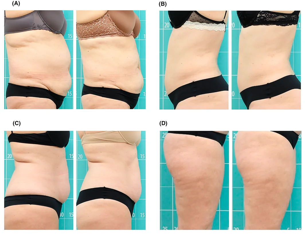
前瞻性单臂研究(西班牙 Asturias 伦理委员会批准),39 例健康女性志愿者(23–68 岁,平均 47 岁,Fitzpatrick II–III),腹部/腰侧 25 例、大
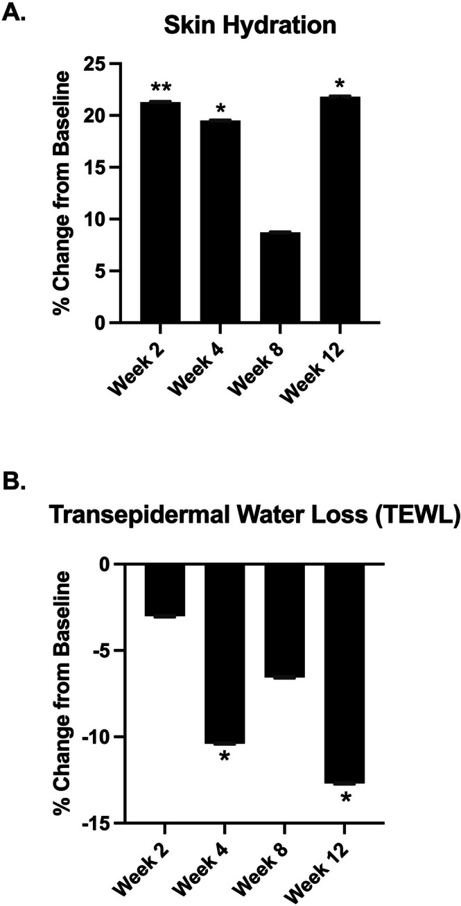
12周前瞻性开放标签单组研究(美国加州):288人筛选、33人入组、29人完成全部访视纳入分析,均为近1年内快速减重≥10%体重者(多为GLP-1/GIP-GLP-1受体激动剂使用