经 Wise 切口的隐蔽 LICAP 皮瓣:用于假体取出后乳腺体积重建的 10 例回顾
2026年10月10日23 min read
Plastic and Reconstructive Surgery Global Open
Plastic and Reconstructive Surgery Global Open
结论与实践要点
本研究为单中心回顾性病例系列,纳入 10 例接受双侧经 Wise 切口 LICAP(外侧肋间动脉穿支)翻转皮瓣行假体取出后乳腺体积重建的患者,平均 BMI 30.84 kg/m²。主要结局为皮瓣成活与 12 个月随访时的形态稳定:10 例皮瓣全部成活,乳头乳晕复合体位置、乳房形态与突度在随访中保持稳定,仅 2 例出现术后血清肿并经随访处理缓解。该术式利用 Wise 切口的横向延长部分在皮下潜行、暴露带蒂脂肪瓣并内翻,以实现“无新切口”的乳腺自体增量。
研究局限明显:样本量仅 10 例,统计效力不足,结论属初步探索性;随访时长有限(12 个月),结局由手术团队主观评估,缺乏客观容积测量与经验证的患者报告结局量表;所有手术由同一团队在同一机构完成,外推性与可重复性受限。该技术适用于有自体脂肪供区、希望避免新增瘢痕且拒绝或不宜再次植入假体的取出后患者;对组织菲薄、脂肪储备不足者并不合适,术前应充分沟通预期。
Background:Selecting the most suitable surgical technique is crucial for successful breast reconstruction after explantation, ensuring optimal aesthetic outcomes and patient satisfaction, and minimizing complications. The authors propose the use of the turnover lateral intercostal artery perforator (LICAP) flap for breast remodeling during reconstructive volumizing procedures in explantation surgery.
Methods:This retrospective case series analyzed patients who underwent LICAP flap procedures for breast remodeling during explantation. Data were collected from medical records, focusing on surgical technique, outcomes, and complications.
Results:The case series included a total of 10 patients who underwent bilateral turnover LICAP flap procedures for breast remodeling during explantation. The average body mass index was 30.84 kg/m 2 . In this study, “scar-free” denotes no new cutaneous incisions beyond those used for the Wise pattern technique; flap harvest was performed through existing access. Flap survival was achieved in all 10 patients. All patients achieved stable results with optimal nipple–areolar complex position, breast shape, and projection during the 12-month follow-up visit. Two patients developed postoperative seromas, which were successfully managed during follow-up appointments.
Conclusions:The turnover LICAP flap is a safe, effective, and reliable option for breast auto-augmentation following breast explantation surgery. This technique demonstrates low donor-site morbidity and complication rates, eliminates the need for patient repositioning, avoids an extended thoracic scar, and provides good aesthetic outcomes.
Question : Are postoperative complications and patient dissatisfaction common after breast explant procedures, and can the turnover lateral intercostal artery perforator (LICAP) flap improve breast remodeling during reconstructive volumizing procedures in explantation surgery?
Findings: This retrospective case series analyzed 10 bilateral patients who underwent turnover LICAP flap procedures for breast remodeling during explantation. Flap survival was attained in all 10 patients. All patients achieved stable results with optimal nipple–areolar complex position, breast shape, and projection during the 1-year follow-up visit.
Meaning: The turnover LICAP flap is a safe, effective, and reliable option for breast auto-augmentation following breast explant surgery.
Surgeons specializing in breast augmentation and reconstruction face an ongoing global challenge with post–breast explant procedures. Postoperative complications, asymmetry, excess skin laxity (ptosis), and patient dissatisfaction can arise from the pressure exerted by the breast implant. This pressure has the potential to cause atrophy or retraction of the parenchymal breast tissue and the underlying pectoral muscle. 1
A frequently observed pathological correlation involves an imbalance between eccentric and concentric forces exerted by the breast implant and the innate elasticity of the skin. This imbalance often manifests as visible tissue atrophy upon explanting the breast prosthesis. Recent publications and social media discussions about autoimmune/autoinflammatory syndrome induced by adjuvants (ASIA), 2 also known as breast implant illness (BII), 3 , 4 and breast implant–associated anaplastic large cell lymphoma 5 , 6 have significantly influenced the perceptions of a large patient population who have previously undergone breast augmentation procedures, leading many patients to seek implant removal and explant reconstruction. 7 – 9
Factors such as aging and implant deterioration often necessitate future interventions, including mastopexy or breast implant exchange procedures. Evolving trends also significantly influence patient decisions regarding desired breast volume and the pursuit of more natural-looking results. These various considerations are crucial to a surgeon’s recommendation for breast implant removal and subsequent reconstructive surgery for eligible patients.
Currently, a variety of autologous tissue flaps based on all pedicles of the breast are being used to enhance and restore breast volume following explantation. The authors propose a novel surgical technique using the lateral intercostal artery perforator (LICAP) flap for breast remodeling during explantation procedures, which avoids extended or unnecessary incisions through the Wise pattern lateral skin undermining. 10 – 15
Initially, the intercostal artery perforator flaps were described and broadly discussed by Hamdi et al. 16 They used them as composite fasciocutaneous flaps with specific angiosomes or cutaneous vascular territories and as rotating flaps. In the past years, modifications in direction, extension, and form of the LICAP flap, as described by Spinelli et al, 17 Yee et al, 18 and Stillaert et al, 19 have been introduced, evidencing the versatility of this flap. Our novel approach introduced the turnover LICAP flap tailored through an extended lateral skin undermining when performing reconstructive volumizing procedures in explantation surgery.
The LICAP flap has been used for oncological patients, breast reconstruction surgery, and cosmetic purposes following significant weight loss or to address lateral breast defects and comorbidities. However, it has never been mentioned as an option for patients who have undergone explantation.
Key advantages of the LICAP flap for different breast reconstruction procedures include its outpatient feasibility, preservation of muscle, reliable blood supply, and minimal donor-site morbidity. The goal is to avoid additional incisions by performing it through an extended lateral skin undermining adjacent to the horizontal pattern of the Wise incision. 17 , 20 – 24
In this article, “scar-free” refers to the absence of additional skin incisions on the breast or lateral chest wall beyond those required for the Wise pattern technique. This term does not imply the absence of internal scar formation, which is inherent to wound healing.
The purpose of this case series was to describe a novel surgical technique using the LICAP flap for breast remodeling during explantation procedures secondary to deficits from prior breast augmentation surgery and provide detailed insights into its application, safety, effectiveness, and potential benefits over traditional methods.
This retrospective case series analyzed patients who underwent LICAP flap procedures for breast remodeling during explantation. Data were collected from medical records, focusing on surgical technique, outcomes, and complications.
Inclusion criteria included (1) patients presenting with any level of mammary gland ptosis; (2) patients for whom, in the opinion of the surgeon, the application of the Wise technique would be clinically appropriate; (3) a body mass Index (BMI) between 20 and 35 kg/m 2 ; (4) patients otherwise in good health; (5) a pinch test in the flap area greater than or equal to 3 cm of thickness; and (6) agreement to the Wise pattern design skin resection. Exclusion criteria include (1) previous liposuction procedures in the axillary and intercostal regions; (2) previous or current diagnosis of ASIA or BII; (3) restrictive pathologies or illnesses; (4) a history of smoking; and (5) previous reconstructive surgery in the past 365 days before surgery.
The study included patients who underwent an LICAP flap for breast reconstruction after explantation. Preoperative images were obtained from all patients. Medical records were retrieved to gather information on patient characteristics, including age, BMI, age of implants, and implant size, and information about the surgical technique. The data were stored in an anonymized manner.
To minimize selection bias, all consecutive patients who met the inclusion criteria during the study period were included. Measurement bias may exist in the estimation of flap volume, as it was calculated based on base width and thickness rather than direct intraoperative volume measurement. Observer bias was reduced by involving at least 2 clinicians in the postoperative evaluation of aesthetic outcomes and complications, using standardized assessment criteria. Given the small sample size, no inferential statistical analyses were performed.
All patients received prophylactic intravenous antibiotics and were positioned in a supine position with sterile dressings.
As described by Jacobs et al, 25 the anterior LICAP flap is a flap based on the perforating arteries, which originate in the coastal segment of the intercostal arteries. It is an axial-pattern flap based on the LICAPs. Using a Doppler probe, at least 2 LICAP pedicles are identified to ensure maximum flap perfusion, and the flap is drawn onto the skin. If visualization is not possible, a 3-cm base pedicle is reserved. The flap has a pivot point at the anterior axillary line. The width of the flap is approximately 10–12 cm, and the posterior limit is the posterior axillary line.
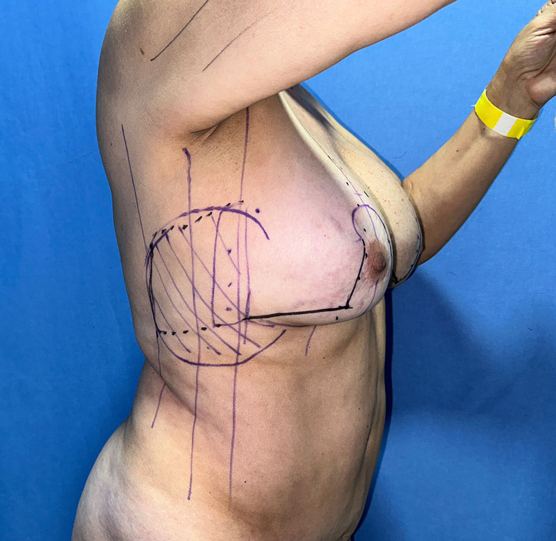
Before surgery, the perforators are identified and marked with Doppler mode using an ultrasound probe. The design of the flap is marked with the patient in a standing position. The incision is made according to the preoperative design and intraoperative modifications (Fig. 1 ).
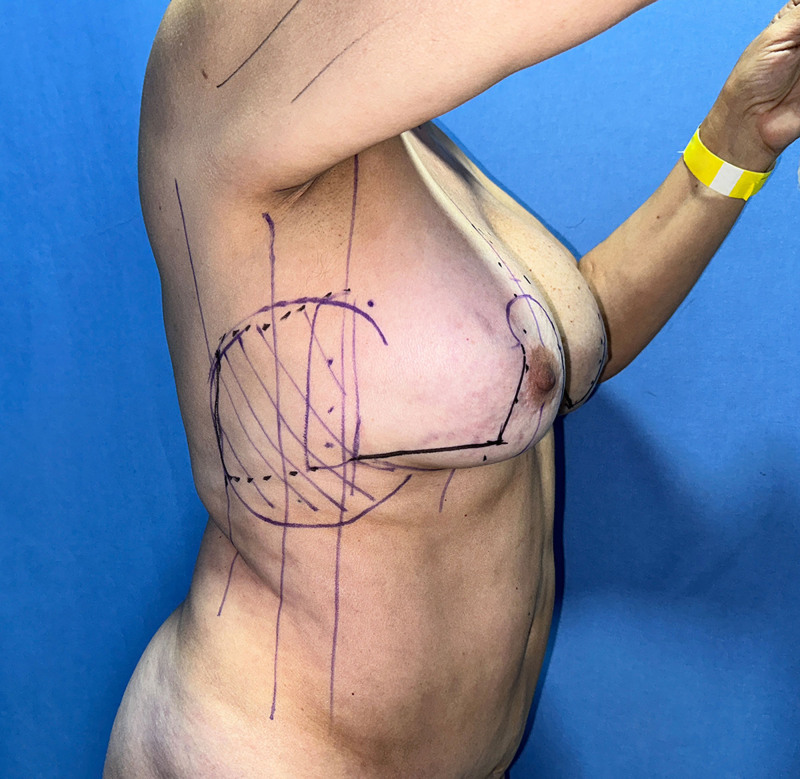
Fig. 1. Surgical description.手术步骤总览示意图。
De-epithelialization is carried out using the Schwartzman maneuver in an inverted Wise pattern to shape the inferior pedicle flap, as described by Ribeiro et al. 26 Following this, the existing breast implant is explanted, and if necessary, the pectoral muscle pocket is closed. The inferior flap is then tailored, elevated, and cephalically fixed. In ideal scenarios, this should be the initial step to avoid compromising or causing further compression of the subsequent LICAP flap.
The LICAP flap is designed in a rectangular shape, adhering to a 3:1 ratio, extending dorsally to the posterior axillary line (Figs. 2 , 3 ). The flap dimensions typically range from a base width of approximately 8 cm to a length of approximately 12 cm (Fig. 4 ). After infiltrating the tissue of the marked area with Klein solution, blunt undermining is performed using Metzenbaum scissors directed toward the skin surface, slightly wider than the flap to be harvested. The average thickness of the skin flap in the donor area was 0.5 cm. One millimeter of methylene blue can be injected subcutaneously adjacent to the previously identified perforators to serve as reference points during the undermining portion of the procedure.
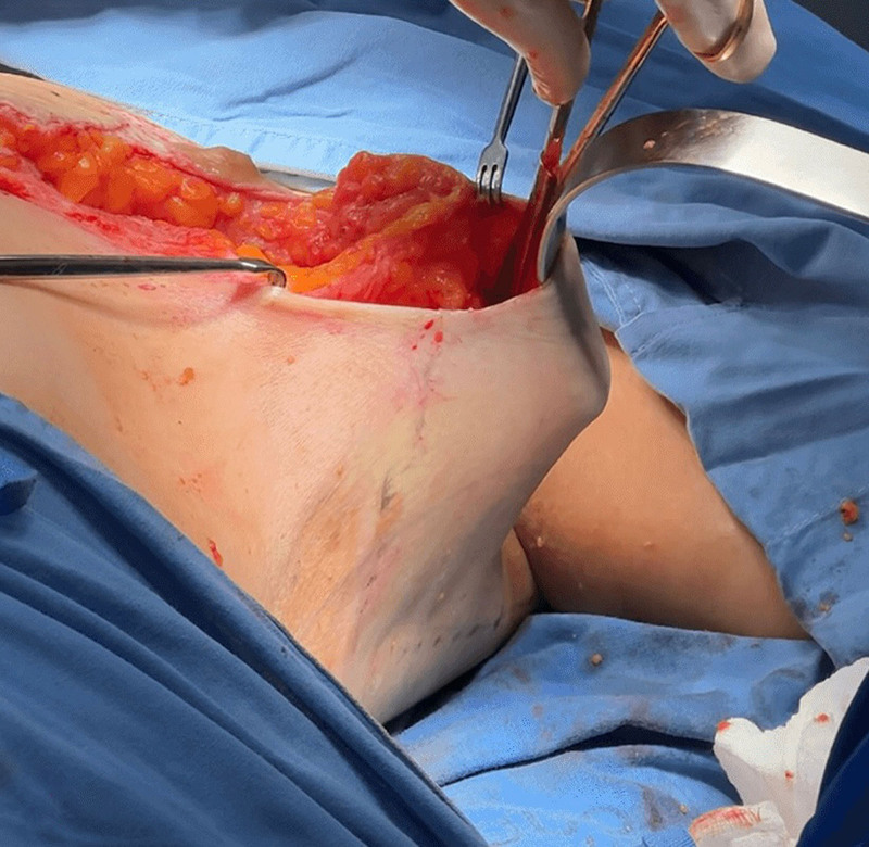
Fig. 2. Preoperative marking.术前体表标记,含 Wise 倒 T 设计。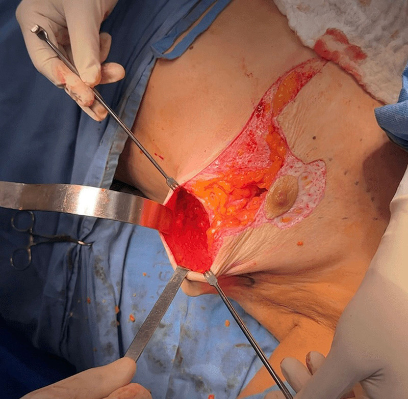
Fig. 3. Turnover LICAP flap tailored through an extended lateral skin undermining. Superior view of the lateral skin undermining, exposing the flap to be elevated and turned over.经外侧皮肤广泛潜行获取的翻转 LICAP 皮瓣(俯视)。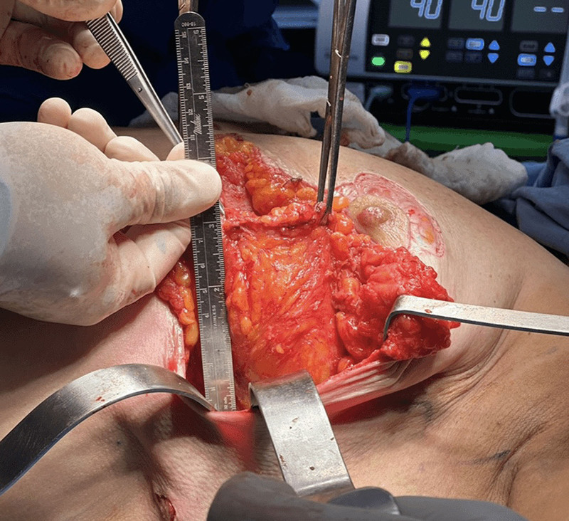
Fig. 4. Tailoring and positioning of the turned-over LICAP flap.翻转 LICAP 皮瓣的塑形与定位。
Electrosurgical dissection is then carried out laterally and dorsally until the latissimus dorsi fascia is identified, reaching premarked points as determined during the preoperative assessment. A minimum of 2 perforators should be included in this zone. The flap is preserved with a pedicle of at least 2 cm in width at its base and is turned over medially. It is sutured in place using 2-0 Vicryl sutures at 3 points: distal end, superior portion, and inferior portion of the flap (Fig. 5 ).
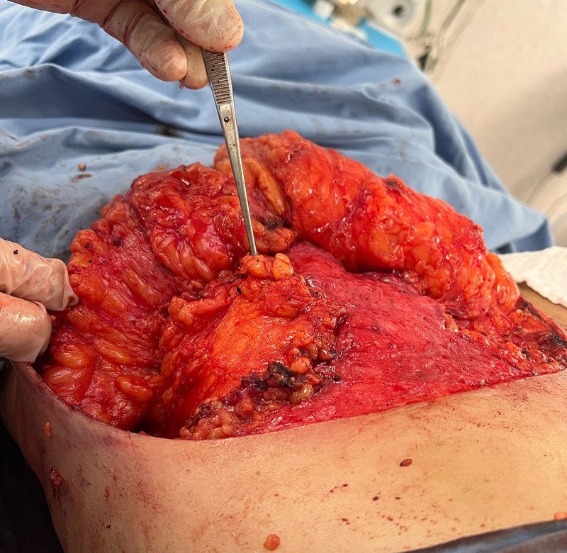
Fig. 5. Overlapping the turned-over LICAP flap on top of the inferior pedicled flap.翻转皮瓣叠加于下蒂皮瓣之上。After identifying the implant plane and having performed the explant, submuscular or dual-plane pockets are closed with 2-0 Vicryl or polydioxanone sutures, without placing a submuscular drain. This is not the case for subglandular pockets, where the pectoralis muscle is preserved.
Strict hemostatic control is maintained at the donor site, and the flap perfusion and viability are confirmed. 27
Subsequent to these steps, the elevation of the inferior pedicle flap and closure of pillars and various planes are executed at the surgeon’s discretion (Fig. 6 ). ( See Video 1 [online] , which displays an animation of the complete procedure.)
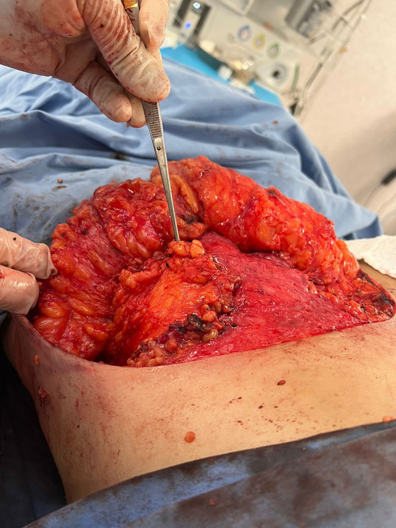
Fig. 6. Intraoperative view of the turnover LICAP flap overlapping the inferior pedicle.术中见翻转 LICAP 皮瓣覆盖下蒂。补充视频 / 附件(原文未随文提供原图,可于出版方页面查看)gox-14-e7636-s001.mp4Video 1. Complete procedure animation.
Ten patients underwent bilateral turnover LICAP flap procedures for postexplant breast volume augmentation. The average BMI was 30.84 kg/m 2 . Patient characteristics are described in Table 1 .
Table 1. Patient Characteristics
| Patients | BMI, kg/m 2 | Patient Age, y | Age of Implants, y | Implant Size (Explanted), cc |
|---|---|---|---|---|
| Patient 1 | 32.1 | 45 | 7 | 355 |
| Patient 2 | 28.7 | 56 | 14 | 325 |
| Patient 3 | 29.3 | 49 | 9 | 375 |
| Patient 4 | 32.4 | 52 | 12 | 400 |
| Patient 5 | 29.8 | 57 | 13 | 380 |
| Patient 6 | 32.5 | 51 | 10 | 350 |
| Patient 7 | 31 | 44 | 11 | 320 |
| Patient 8 | 30 | 50 | 12 | 400 |
| Patient 9 | 32.7 | 53 | 15 | 450 |
| Patient 10 | 30 | 47 | 16 | 420 |
The case series included a total of 10 bilateral patients who underwent turnover LICAP flap procedures for breast remodeling during explantation. Pre- and postoperative views for cases 1 and 2 are shown in Figures 7 – 14 .
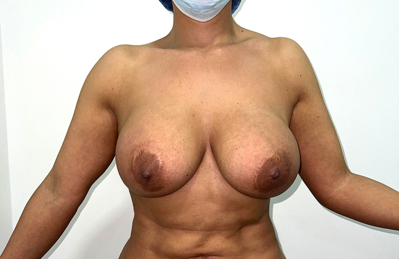
Fig. 7. Preoperative frontal view.病例 1 术前正面观。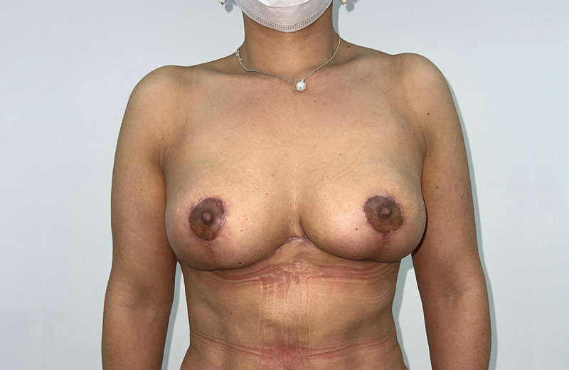
Fig. 8. Postoperative frontal view at 9-month follow-up.病例 1 术后 9 个月正面观。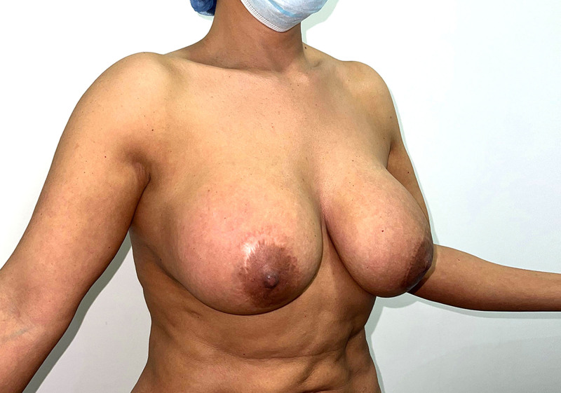
Fig. 9. Preoperative oblique view.病例 1 术前斜位观。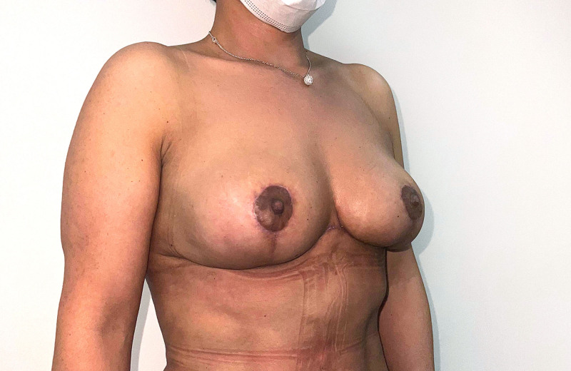
Fig. 10. Postoperative oblique view at 9-month follow-up.病例 1 术后 9 个月斜位观。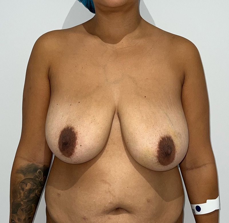
Fig. 11. Preoperative frontal view.病例 2 术前正面观。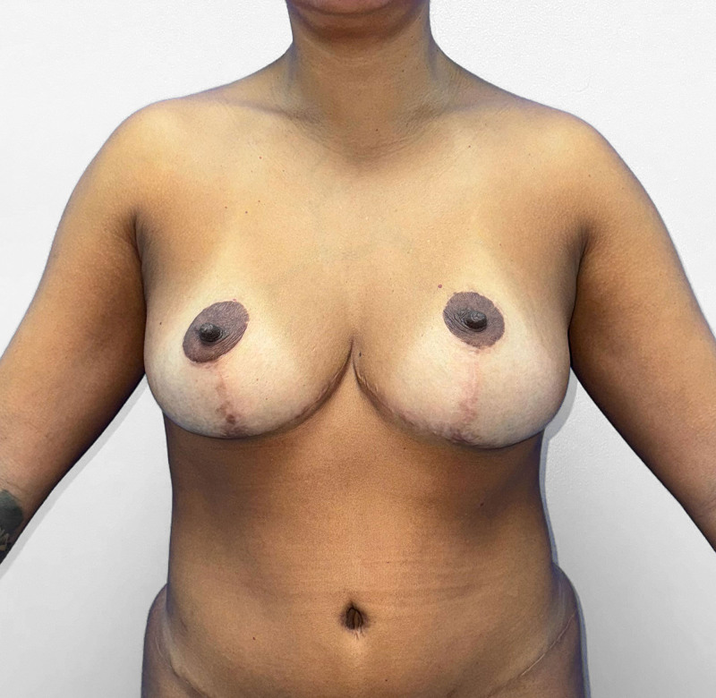
Fig. 12. Postoperative frontal view at 12-month follow-up.病例 2 术后 12 个月正面观。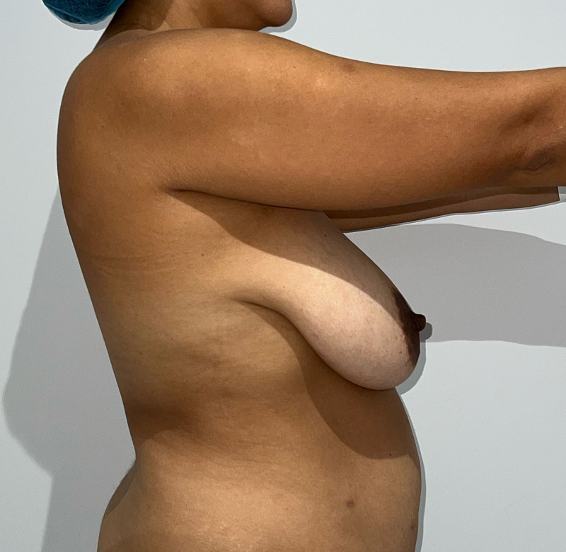
Fig. 13. Clinical image, case 2: preoperative right profile view.病例 2 术前右侧侧位观。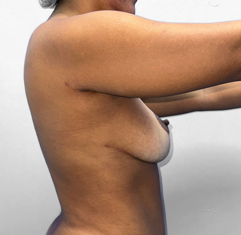
Fig. 14. Postoperative right profile view at 12-month follow-up.病例 2 术后 12 个月右侧侧位观。
Flap survival was achieved in all 10 patients. An intraoperatively inserted Jackson-Pratt drain was used to manage the accumulation of postoperative serosanguineous fluid. No circulation problems or sagging of skin were observed on the flap donor site.
At the 1-month postoperative follow-up, all patients achieved stable results with optimal nipple–areolar complex position, breast shape, and projection. A depression in the donor area was observed in all patients, but it did not cause any patient dissatisfaction (Fig. 15 ). Instead, it seemed to satisfy most patients, as it narrowed the thorax diameter, creating a slimmer appearance and enhancing the contour of the lateral aspect of the breasts. All cases completed a postoperative 12-month follow-up.
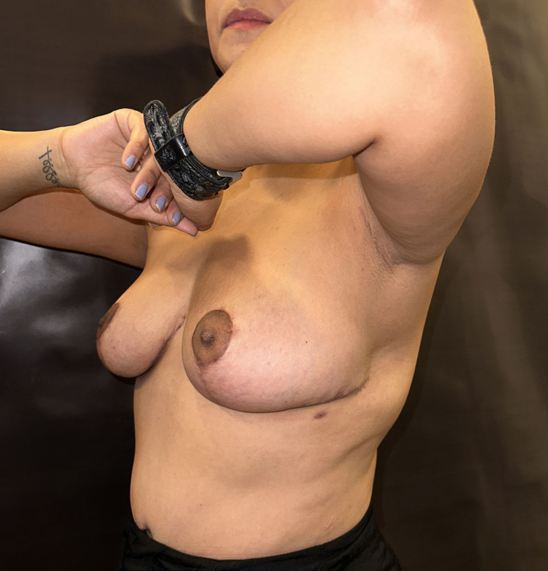
Fig. 15. Lateral view of the donor-site depression postsurgery at 12 months, shown side by side.术后 12 个月供区凹陷侧位对照。
Postoperative complications were retrieved from medical records up to 1 month after the surgery. A complication was defined as any issue at the recipient or donor site, including wound dehiscence, infection, seroma, fat necrosis, reexploration, and flap loss. Two patients developed postoperative seromas, which were managed during their follow-up appointments. ( See Video 2 [online] , which displays the seroma drainage and the postoperative view at 10 days follow-up.) In case 2 (Figs. 11 – 14 ), there was a visible dropout asymmetry, possibly due to insufficient skin resection, not related to the flap viability.
补充视频 / 附件(原文未随文提供原图,可于出版方页面查看)gox-14-e7636-s002.mp4Video 2. Seroma drainage. Postoperative, 10 days follow-up.
Breast reconstruction after atrophy caused by long-term breast implants remains a challenge for surgeons, especially when managing patient expectations. The primary purpose of the initial consultation with a patient seeking breast implant explantation should be to diagnose, understand, and evaluate the patient thoroughly to determine the genuine need for the procedure. Selecting the most suitable surgical technique is crucial for successful breast reconstruction after explantation, ensuring optimal aesthetic outcomes and patient satisfaction, and minimizing complications. Although we have observed an increase in consultations for explantation related to ASIA, also referred to as BII, and breast implant–associated anaplastic large cell lymphoma, in our practice, the number of procedures driven by these concerns does not exceed 10% of consultations.
The surgeon’s role is to diagnose, educate, and offer options. The vast majority of consultations seem to be motivated by anxiety and fear rather than by objective findings, facts, or florid or specific symptoms. In most cases, we are able to rule out this condition, counsel patients against unnecessary explantation, or perform a revision procedure such as prosthesis replacement, mastopexy, or pocket adjustment.
Imaging studies such as ultrasound and magnetic resonance imaging, together with antibody testing and rheumatology consultation, are recommended when an abnormality or autoimmune disease is suspected; however, no positive findings have been documented in these evaluations. Although we have not diagnosed autoimmune disease in any case, the surgeon must remain vigilant for symptoms that may arise from silicone infiltration into the breast parenchyma.
For this reason, we highlight this condition in the present article. Our purpose is to raise awareness of the syndromes, conditions, and malignancies that have been documented and recognized in the scientific literature.
Although the LICAP flap is well established in the literature, its application in healthy patients seeking cosmetic benefits has been limited by the potential for extended scarring in the axillary region, a drawback particularly associated with the propeller approach 16 and traditional turnover methods. 28 These existing approaches are shown in Video 3. 29 ( See Video 3 [online] , which displays the existing techniques using the LICAP flap.)
However, its application in postexplantation breast volume restoration has not previously been reported. Our technique takes advantage of the lateral extension of the horizontal scar in the inverted Wise pattern, undermining the skin and exposing the pedicled fat flap, which is turned medially to augment breast volume. While marking the patient, the surgeon can map the perforating intercostal arteries that will supply blood flow to the flap.
A key innovation of this new technique is the ability to elevate the flap through the lower incision of the inverted Wise pattern, allowing for flap exposure in the supine position without the need for patient repositioning in surgery. This approach effectively creates a “scar-free” turnover LICAP flap by enabling axillary skin undermining and eliminating new axillary incisions. These distinctive features make the technique an attractive and viable option for patients undergoing postexplant breast reconstruction.
Our case series demonstrated the versatility of LICAP flaps in addressing defects that may arise during explantation procedures. Initial clinical outcomes with this novel procedure have been positive. Local perforator flaps provide a robust solution for patients with significant volume or contour deficits after explantation. Local flap reconstruction, including the LICAP flap, is associated with minimal donor-site morbidity and a lower complication rate compared with more extensive reconstructive techniques.
This study is limited by a small sample size (n = 10), restricting statistical power and rendering the findings preliminary and exploratory rather than definitive. Outcomes were short term and assessed subjectively by the surgical team, without objective volumetric methods or validated patient-reported outcome measures. Furthermore, all procedures were performed by a single surgical team at 1 institution, which may reduce reproducibility and limit generalizability to other clinical settings.
A representative flap with an 8-cm base, 12-cm length, and 2- to 2.5-cm thickness can recruit roughly 200 cm 3 of tissue, depending on patients’ characteristics. The main value of the flap lies not in its standalone volume but in its synergy with the inferior pedicle, together producing superior lateral breast shaping.
In individuals with a BMI greater than 30 kg/m 2 , breast tissue is usually abundant, although not invariably so. The technique described here targets patients with breast atrophy in whom the inferior pedicle alone may be insufficient. The turnover LICAP flap does not replace the inferior pedicle; it complements it, improving lateral contour when overlaid on the lower pedicle. A BMI of 30 kg/m 2 or greater is required to ensure that the donor site contains adequate tissue.
When available, an ultrasound probe can locate perforators from the lateral intercostal arteries in the fifth and seventh intercostal spaces along the mid-axillary line. A standard fasciocutaneous propeller flap usually includes a single artery with its venae comitantes. In the turnover approach presented here, the flap’s vertical base allows incorporation of a second intercostal perforator without ultrasound guidance. The LICAP flap has been used successfully for decades to address lateral breast defects. This technique has a short learning curve and offers a change in the form, shape, and dissection technique through skin undermining, preserving the same principle of the conventional fasciocutaneous LICAP flap techniques that exist today.
Our case series demonstrated the versatility of LICAP flaps in addressing defects that may arise during explantation procedures. Initial clinical outcomes with this novel procedure have been overwhelmingly positive. Local perforator flaps provide a robust solution for patients with significant volume or contour deficits after explantation. Local flap reconstruction, including the LICAP flap, is associated with minimal donor-site morbidity and a lower complication rate compared with more extensive reconstructive techniques. A depression in the donor area secondary to the procedure seems to satisfy most patients, creating a slimmer appearance and enhancing the contour of the lateral aspect of the breasts.
The turnover LICAP flap requires proficient dissection of lateral perforators, with a risk of vascular compromise if not carefully performed. Its use may be limited in patients with low BMI or insufficient lateral thoracic tissue. Cosmetic results can be unpredictable, especially in cases of significant atrophy or challenging anatomy, necessitating careful patient selection and expectation management. A depression in the donor area secondary to the procedure seems to satisfy most patients, creating a slimmer appearance and enhancing the contour of the lateral aspect of the breasts. Although initial experience indicates that the technique is accessible, studies in larger and more varied cohorts are needed to clarify the learning curve and consistency across different surgical teams.
Internal validity is limited by subjective outcome assessment, as breast volume and cosmetic changes were evaluated only through clinical judgment by 2 unblinded clinicians, without imaging or standardized measurements, increasing the risk of observer and measurement bias. External validity is also constrained because all cases were performed at a single center by 1 team within a small cohort, limiting applicability to broader populations and diverse clinical environments. Consequently, these preliminary observations should be interpreted with appropriate caution.
补充视频 / 附件(原文未随文提供原图,可于出版方页面查看)gox-14-e7636-s003.mp4Video 3. Animated video showing the existing techniques using the LICAP flap.
Our initial experience with the turnover LICAP flap demonstrated a safe, effective, and reliable option for breast auto-augmentation following breast explant surgery. It offers an alternative option for patients with significant volume or contour deficits after explantation.
Our case series experienced low donor-site morbidity and complication rates. The scarless turnover LICAP flap eliminated the need for patient repositioning during surgery, while avoiding an extended thoracic scar and providing good aesthetic outcomes.
The authors have no financial interest to declare in relation to the content of this article.
Informed consent for surgical procedures and the use of clinical images was obtained from all subjects included in the case series.
Artificial intelligence was used only to improve grammar and syntax. It was not used to generate the content of the article.
声明:中文精读 · 仅供学术参考。内容来自公开文献检索,不代表本人观点,不构成诊疗建议。 医疗美容需在正规医疗机构由执业医师实施。
基于相同主题推荐 · 继续深挖
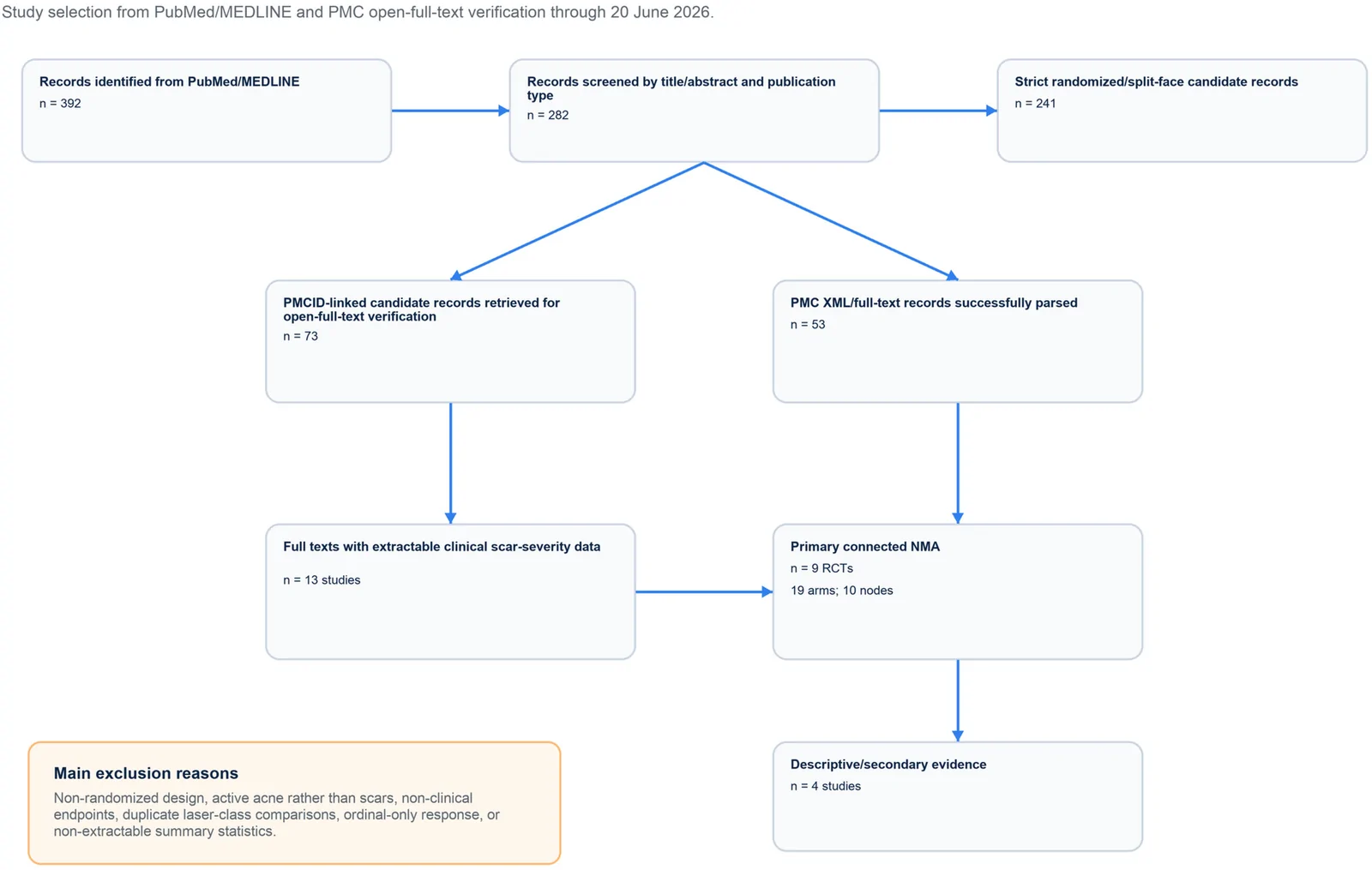
PROSPERO注册的系统综述,纳入9项随机/分脸随机试验、19个治疗臂,构成稀疏连接网络并采用频率学随机效应网络荟萃分析。
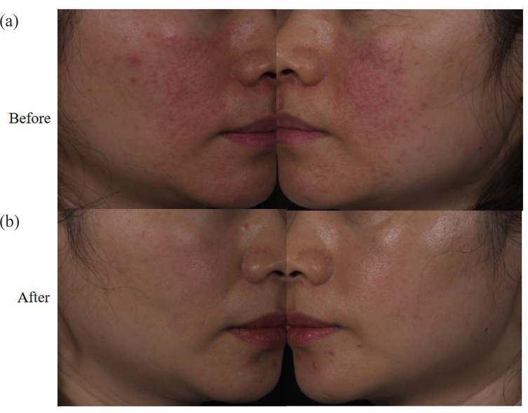
回顾性纳入63例红斑毛细血管扩张型玫瑰痤疮(ETR)中国患者,均采用555–950 nm与530–750 nm双滤光片窄谱IPL治疗。
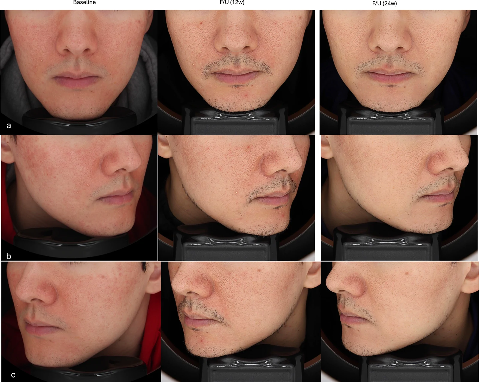
真实世界回顾性队列纳入50例临床诊断玫瑰痤疮成人,80%为女性、中位年龄33岁,多数(76%)为Fitzpatrick III型肤色。