胶原刺激类填充剂异物肉芽肿(FBG):40 项研究 117 例系统综述
2026年10月15日23 min read
Journal of Cosmetic Dermatology
结论与实践要点
该综述的价值在于把「罕见但棘手」的迟发性异物肉芽肿做了系统性汇总,可直接支撑术前沟通与并发症处置预案。几个可落地的要点:① 潜伏期认知——中位 20 个月、最长 15 年,术后短期无异常不能排除迟发反应,随访宣教应覆盖「数年」尺度;② 部位意识——口周与眶周为高发区,与面部肌肉频繁活动、颗粒迁移有关,在这些区域使用微球类产品应更保守;③ 材料差异——不可降解 PMMA 的报告例数与潜伏期高于可降解材料,CaHA/PCL 类潜伏期相对短,提示降解性能影响反应时程;④ 治疗现实——激素病灶内注射为一线常用手段,但顽固者依赖手术切除且难以切净,预防(正规产品、恰当层次、不过量、避免炎性环境下注射)比治疗更关键。
证据边界须明示:纳入研究以病例报告/系列为主(39/40),无分母暴露人群,构成比不能等同于真实发生率,只能反映「报告频率」;缺乏标准化 FBG 定义与组织学确认是主要偏倚来源。作者另指出注射相关生物膜、复溶产品现场配制污染、真皮内注射免疫反应性更高等机制可能与 FBG 相关,提示规范化无菌操作与层次选择的临床意义。
BackgroundCollagen‐stimulating filler has boomed as a new welcoming option in aesthetic treatments by activating the synthesis of endogenous collagen. However, hyperactive or inappropriate foreign‐body stimulatory reactions may trigger intractable foreign body granulomas (FBGs). A comprehensive understanding of the FBGs associated with the augmenting effect of collagen biostimulants can help clinicians perform treatments in a safer and more effective way.
MethodsA literature search was conducted in Web of Science, PubMed, Embase, and Scopus to identify relevant clinical reports about the FBGs induced by six types of commercially available collagen stimulators up to August 2024.
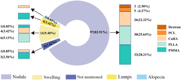
ResultsA total of 40 studies involving 117 patients were included. All but eight cases were women, and the mean age was 52.63 years (range 29–78 years). The most reported cases of FBGs were associated with PMMA (35.04%), followed by PLLA (30.77%), CaHA (27.35%), PCL (4.27%), and dextran‐based filler (2.56%). The time until initial detection of FBGs reaction ranged from 1 week to 15 years (mean 20.18 months). Nodules were the most frequent complication (82.91%), followed by swelling (9.40%) and lumps (3.42%). The perioral region was the reported high‐risk region for FBGs. Intralesional injection of steroids was performed most frequently (21.37%), but symptoms were often resolved through surgical removal.
ConclusionsAll included collagen biostimulators can induce FBGs reactions, with the nondegradable fillers having a higher reported incidence and a longer latency period. Timely recognition, diagnosis, and appropriate management of FBGs behind tissue augmentation with collagen stimulator filler are necessary for clinicians to optimize efficacy and minimize adverse events.
Level of EvidenceV.
Keywords: collagen stimulators、foreign body granuloma、foreign body reactions、treatment recommendations
With the increased demand for facial rejuvenation and a shift toward a long‐term restoration effect, collagen stimulatory fillers for facial augmentation have dramatically gained popularity as an alternative to traditional volume‐based procedures, such as the injection of hyaluronic acid (HA) derivatives. These approved synthetic fillers are prominent representatives of collagen stimulants, such as polymethyl methacrylate (PMMA), polyvinyl alcohol (PVOH), polylactic acid (PLLA), polycaprolactone (PCL), calcium hydroxyapatite (CaHA), and dextran containing fillers (Table S1 ). As an injectable dermal fillers, they were formulated into microspheres or particles and suspended in different kinds of carrier medium. These biostimulatory fillers provide initial volume replacement as well as the late additional neocollagenesis for sustained facial volumization and skin quality. Several lines of evidence suggest that the biological mechanisms behind the augmenting effect most likely depend on the subclinical foreign body tissue reaction of the filler, mediated by moderate inflammatory cell infiltration, foreign body cell recognition, fibroblast growth, material encapsulation, and the consequent collagen deposition. Notably, these synthetic particulate materials, as potential foreign bodies, may also clinically cause a series of rare foreign body reactions (FBRs) during the intended process of promoting collagen regeneration, such as hypopigmentation, lumps, nodular masses, hypertrophic scars, localized swelling, fibrosis, severe skin necrosis, chronic infections, or even serious autoimmune diseases [ 1 , 2 ]. These semipermanent or permanent fillers were more likely to be associated with lumps and nodules than other complications, generally occurring late (> 1 month) [ 3 ]. Furthermore, certain material characteristics may play a vital role in bringing about microsphere‐containing fillers more prone to these FBRs than others. Foreign body granuloma (FBGs) is the main manifestation of chronic FBRs and is classified as a grade 4 FBR. The classification of FBRs described by Duranti et al. shows the classic histopathological features of foreign body granulomas (FBGs), that is, granulomas with encapsulated implants and clear foreign body reactions [ 4 ]. Lemperle G's comprehensive review concluded that the FBGs of particulate injectables typically present as sclerosing granulomas, and the possibility of a blend of cystic or edematous granulomas may also exist [ 5 ].
Studies of the FBRs of particulate fillers such as these suggested that FBGs are a rare, intractable, and recurrent presentation that requires immediate attention [ 6 , 7 , 8 , 9 ]. A particularly challenging problem is that permanent fillers often lack effective solvents, leading to complications that are typically persistent. Data from several studies suggested that the incidence of complications in nonabsorbable dermal fillers was nearly 10 times higher than that of absorbable fillers [ 10 , 11 , 12 ]. In addition, permanent products, like PMMA and PVOH fillers, are more likely to be the origin of unaesthetic, recurrent, and invalidating FBGs that are often very long‐lasting and resistant to various local treatments [ 13 ]. However, many of biomolecular and clinical studies up to now have been extensively focusing on the augmenting effect of stimulatory filler. There remains a paucity of evidence on the long‐term safety, especially the nonbiodegradable stimulatory filler. Herein, we present the first systematic review and general discussion regarding to the status and management of FBGs related to the collagen stimulants to better guide practitioners in facilitating safer clinical procedures.
1
The search strategy for this study was guided by the Population, Intervention, Comparison, Outcome (PICO) framework [ 14 ]. The detailed Medical Subject Headings (MeSH) terms retrieved and search strategies for this research were shown in Appendix S1 . The fillers retrieved above were as shown in Table S1 . An extensive literature search was performed by three reviewers (H.Q., H.W., and S.L.) via Web of Science, PubMed, Embase, and Scopus in August 2024 to identify all reports pertaining to FBGs of the PCL, PLLA, CaHA, PVOH, PMMA, and dextran‐based regenerative biostimulators after cosmetic procedures. FBGs reaction induced by fillers was mainly diagnosed by clinical manifestation and pathologic examination. Two investigators (C.Lo. and Q.J.) independently conducted initial screening on abstracts and full‐text reviews of relevant studies according to the predetermined criteria (Figure 1 ). Any disagreements or ambiguities with the search results and data analysis were adjudicated by discussion with the senior author (DM.W.).
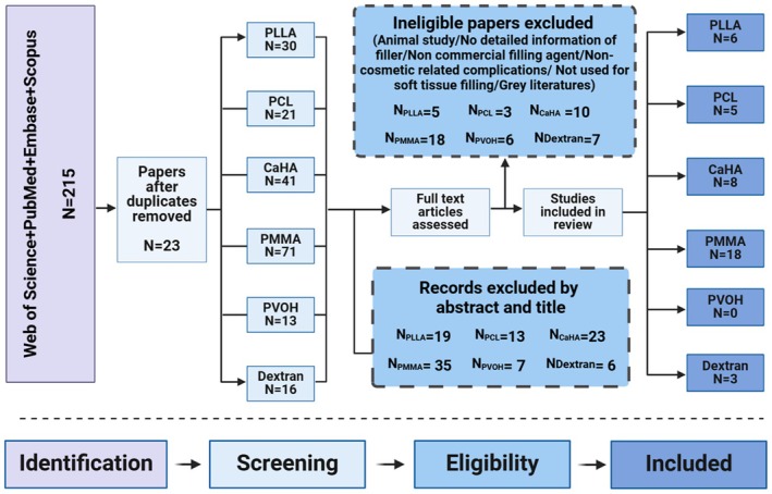
FIGURE 1 The flow diagram of the literature screening and study selection process.文献筛选流程(PRISMA):215 篇检索记录经去重、筛选后 40 篇纳入。2
A total of 215 articles were retrieved with the prespecified search criteria. Following the removal of duplicates, 192 studies were screened, and 89 articles underwent full‐text assessment (Figure 1 ). Forty studies were finally included in this systematic review. The patient clinical characteristics, types of treatments implemented, and prognosis of the included studies are detailedly presented in Table S2 .
2.1
Thirty‐nine case reports and one retrospective study describing a total of 117 patients who suffered from FBGs after receiving injections met final inclusion criteria. All 117 patients, aged 29–78 years (median, 52.63 years), were 109 women (93.16%) and 8 men (6.84%). As seen in Figure 2 , PMMA (35.04%) has been reported to cause the most granulomas reactions, followed by PLLA (30.77%) and CaHA (27.35%). This result was closely related to the properties of the materials, such as immunogenicity and degradability. Six different types of dermal fillers and nine products were reported in these patients, with Artecoll (Rofil Medical, Breda, Netherlands) ( n = 35/29.91%) and Radiesse (Merz Pharmaceuticals, Frankfurt, Germany) ( n = 32/27.35%) being the most reported. Newfill (Sanofi Aventis, Paris, France) was reported in 20 patients (17.09%) and other fillers were also described, such as Sculptra (also known as New‐Fill; Sanofi Aventis, Paris, France), ELLANSÉ (Aqtis Medical, Utrecht, Netherlands), Matridex (BioPolymer, Samedan, Switzerland), Bioplasty (Silikon Industria, Rio de Janeiro, Brazil), and Metacrill (Nutricell, Campinas, Brazil).
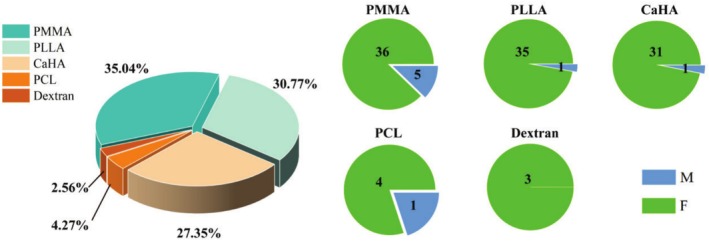
FIGURE 2 Number of patients with FBGs due to various microspheres‐containing dermal fillers (F: female; M: male).各微球类填充剂所致 FBG 患者例数分布。The time elapsed from injection to initial detection of the lesion ranged from 1 week to 15 years, with a median of 20.18 months. The time until complications varied by filler type: the median time was 18.75 months for PLLA, 13.20 months for PCL, 7.35 months for CaHA, 35.34 months for PMMA, and 2.33 months for dextran microspheres (Figure 3 ). A longitudinal study of the human histology and persistence of dextran microspheres by Lemperle G reported that early redness and swelling after implantation can occur within 10 days and may persist for up to 3 months [ 53 ]. This was consistent with the reported window period (range: 1–5 months) for FBRs of dextran microspheres and is also one of the reasons for the earliest occurrence of granuloma formation in these six fillers. As an inert implant, FBGs associated with PMMA often appeared later, sometimes as long as 15 years after initial injection, and could persist the longest.
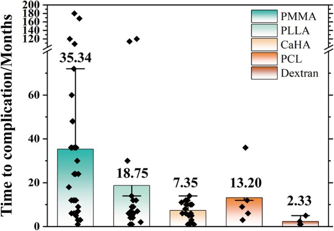
FIGURE 3 Months between filler injection and FBG's first development.注射至 FBG 首发的时间分布(按填充剂类型)。2.2
The granulomatous lesions at presentation appeared from lumps or tumefaction to the most common nodules, occasionally with migration occurrence or necrosis manifestations (Figure 4 ). Among the affected regions, the oral and perioral areas were the most common anatomical locations of granulomas, accounting for 30.77% of cases. This includes the labial mucosa, labial vestibule, and mental area. Other notable sites represented the common affected anatomical locations of granulomas. The lips represent 8.55% of cases, the glabellar area was 5.13%, and the periocular area was 4.27% (Figure 5 ). Among the multiple lesion sites, in addition to the conventional facial injection areas, other parts of the body were also involved, such as the buttocks and thighs. Occasional reports also mention the hands, jawline, and neck.
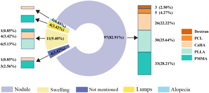
FIGURE 4 The clinical manifestations and proportions of granuloma reactions for each filler.各填充剂肉芽肿反应的临床表现与占比。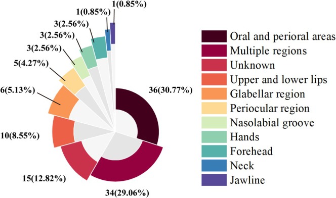
FIGURE 5 The reported sites and proportions of granuloma reactions' occurrence.FBG 发生部位与占比。2.3
As is shown in Figure 6 , the majority of the cases were managed by intralesional injections of steroids (25/21.37%), followed by surgical removal of lesions (18/15.38%), laser treatment (5/4.27%), and antibiotics (2/1.71%). Furthermore, allopurinol, methotrexate, and fluorouracil were also reported in other cases. However, the treatment protocol was not mentioned in 48 cases, 1 case was lost to follow‐up, and 6 cases did not receive any intervention.
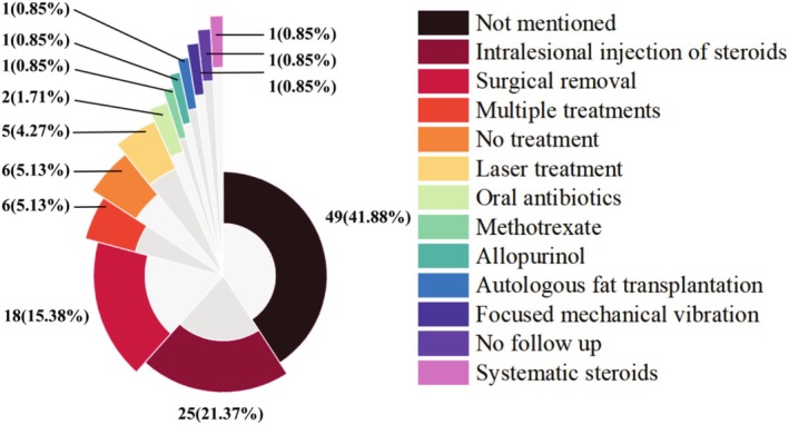
FIGURE 6 The treatment modalities of the included cases.已报告病例采用的治疗方式分布。2.4
Figure 7 illustrated that there was remission of the nodule (27.35%) or complete resolution (10.26%). Eight cases reported persistent lesions with no significant improvement in lesion size or symptoms. A total of 65 cases were lost to follow‐up or did not report on the outcome. It is worth noting that most refractory cases are mainly resolved through surgical excision.
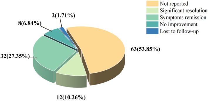
FIGURE 7 The treatment prognosis of the included cases.治疗结局分布(缓解/完全消退/持续存在/失访)。3
Microsphere‐based tissue stimulators were used more as a sculpting and regenerative agent rather than just a volumizing filler. These fillers are considered relatively safe, with most adverse reactions occurring either immediately or shortly after injection. Noteworthy, the long‐term or delayed‐onset adverse effects caused by any implanted medical device are often considered FBRs, which tend to occur several months to years after initial injection and usually form characteristic FBGs microscopically. Ideally, the limited FBRs around the injected material are the body's natural response to the implant. That is often presented as a monolayer of macrophages or fibrocytes surrounding microspheres and enveloped by a zone of new fibrous tissue, which is the true histologic basis of the volume restoration and not a fibrosis reaction. However, variable degrees of fibrosis with an accumulation of product were often noted in the manifestation of granulomatous FBR, especially induced by nonbiodegradable microsphere filling agents. This accumulation of tissue regeneration differs in that it is primarily due to the host tissue's excessive reaction to small amounts of the product [ 54 ]. As Werschler put it, the granulomatous reaction was not identical to the clinical biostimulator‐induced nodules, especially in the primary mechanism of action, histopathologic response, and even treatment approaches [ 55 ]. It can be seen in Table S1 that the main histological appearance of FBGs associated with collagen biostimulators varied from fillers to fillers, mainly depending on the chemical nature of the microspheres, their surface structure, and the presence of carriers. Knowing the histological characterization of FBGs is important for our increased understanding and distinguishing of different injected substances.
It has previously been observed that microsphere size was among the most important factors for FBRs. Most of these particle‐containing fillers are presented with a diameter of 30–50 μm to resist macrophage phagocytosis, but they still cannot completely avoid a series of potential FBRs, not to mention the potential impact of smaller particle size, irregular shape, rough surface, inappropriate content, and surface charge. As summarized in our review, the FBGs caused by the second‐generation PMMA filler (Artecoll), containing a higher volume of particles (< 20 μm in diameter), are significantly higher than those reported for Artefill. Another PMMA‐based product, Metacrill, may also suffer from late‐onset granulomas due to a wide microsphere size distribution (1–80 μm). Similarly, FBRs in PLLA fillers mainly occurred in the Sculptra product, which has more uniform particle sizes than those of Newfill. As would be expected, implantation with irregular surface microspheres (Bioplastique (Bioplasty, St. Paul, Minnesota), Dermalive (Dermatech, Paris, France)) is prone to have a higher probability of inducing FBRs compared to smoothly surfaced microspheres [ 54 ]. Notably, the reported cases of FBGs to PMMA ( n = 41/35.04%) in our study are more than the cases of HA ( n = 41/28.80%, reported in 2023). Nondegradable filler is often considered as latent “living implants” to exert long‐term stimulants for connective tissue formation. This also accords with our observations, which showed that the incidence rate of granuloma formation was higher for nondegradable fillers compared to resorbable fillers in our study. As reported, the occurrence of granuloma from PMMA can happen as long as 15 years after postinjection.
Cellulose is a common carrier for injectable microsphere materials, such as PVOH, PLLA, PCL, and CaHA. CaHA and PLLA can also use cross‐linked hyaluronic acid (HA) as an excipient. PMMA‐based injectable products usually use bovine collagen as the carrier. To some extent, the source, degradation, and by‐products of the carrier material can also induce a host response. The bovine collagen carrier in PMMA fillers may trigger allergic reactions, which could also be a potential risk factor for causing FBRs. Some authors also suggested that, except for the type of carrier itself, the biological structure of the excipient (viscosity, morphology, cohesiveness, hardness, hydrophilia, surface charge, and roughness) or processing techniques (cross‐linking form) may also be other factors influencing the outcomes of fillers [ 56 ]. Therefore, some inert and asymptomatic complications arising from the above inherent material factor of fillers tend to lean more toward noninflammatory, nonspecific immune responses. Although manufacturers often claim that these fillers have good tolerance, are nonimmunogenic, or that specific immune complications are very rare, we cannot ignore the possibility of such reactions.
Further findings revealed that the occurrence of FBGs was well influenced by the site and location of injection. As concluded by Daley et al., the migration of the particles appears to be linked to the formation of ectopic granuloma lesions, especially in areas with frequent facial movements, such as the perioral area, eyelids, lips, neck, and cheeks [ 15 , 57 ].
Given the off‐label use of these fillers, the locations where FBG occurs in clinical practice are more variable than the lesion sites reported in existing literature. Evidence indicates that the dermis exhibits heightened immune reactivity, leading to an increased propensity for granuloma formation following intradermal injections compared to subcutaneous or dermal‐subcutaneous junction administration. These lyophilized powder products, such as fillers containing PLLA and CaHA, often require on‐site preparation before injection. The carrier matrix and microspheres typically need to be blended or dispersed in sterile distilled water or saline to obtain a homogeneous mixture. Therefore, too superficial an injection, a high concentration of microspheres within the filler, a lack of massage, an excessive injection dose, insufficient intervals, incorrect product preparation, or hydration time may also lead to the focal aggregation of microspheres and then the induced nonimmune reactions, such as the asymptomatic nodule [ 44 ].
Our review presented the finding that the nodules in reported complications were mostly characterized as inflammatory granulomas. Many studies examined that bacteria inoculation during injection through contiguous direct extension or hematological spread may form biofilms. This can resist bodies and external degradation measures on the filler materials [ 58 , 59 , 60 ]. With polysaccharide‐rich heterogeneous structures embedding various microorganisms, these biofilms can evade the immune system and effectively inhibit macrophage phagocytosis. Studies have shown that these biofilms often act as bacterial antibiotic resistance and tolerance [ 61 , 62 , 63 ]. This may be one of the reliable explanations for the delayed foreign body reactions, such as persistent nodule granulomas. Additionally, Lemperle et al. have concluded that there may be a possible correlation between incidents provoking alterations in immune dynamics, like facial trauma, face‐lift operation, systemic infections, flu‐like syndrome, and herpes labialis, and FBGs before their clinical onset [ 5 ]. Some researchers stated certain medications, such as interferon‐α and ribavirin, that may induce skin sarcoidosis can act as a triggering cause for the occurrence of granulomatous reactions of the preexisting filler [ 38 ]. Exceptionally sensitive conditions or suffering from autoimmune diseases may also be an increased predisposing factor in FBRs causation. It seems possible that these hosts’ immune statuses or the interaction with immunostimulatory medications may stimulate the memory of macrophages and prompt a new attack, especially in cases in which filler is residual or a second injection [ 37 , 44 ]. In this case, there is a probability that FBGs have cyclic periods of activation and remission in clinic. Collectively, further studies need to be conducted to elucidate the mechanisms that cause late‐onset granuloma formation.
The timing and progress of management actions are fundamental to the outcomes of resolve, remission, and exacerbation. These products that require reconstitution with excipients may cause early to immediate‐stage nodules after injection, which could be the result of particle accumulation rather than a granulomatous reaction. Surveys such as that conducted by McCarthy have shown that focused mechanical vibration relying on the combination of aqueous diluent and microneedling vibration seemed to be a promising strategy for the rapid reversal of noninflammatory nodules resulting from CaHA‐based injections [ 45 ]. For such nodules, manual dispersion of particles like local massage, mechanical vibration, or dilute saline can be considered as treatment options. There were also studies reporting that for such nodules, symptoms may improve even without any intervention by the spontaneous biodegradation of the filler [ 18 ].
As for some later‐stage ones, being managed with intralesional steroid, 5‐fluorouracil, methotrexate, bleomycin, and prednisone injections, or combined with oral agents (minocycline, allopurinol, colchicine, cyclosporine, corticosteroids, methotrexate, and doxycycline), and energy‐based devices (LED, erbium laser, intense pulsed light, fractionated laser, CO 2 laser, and microfocused ultrasound) have been typically suggested as the symptomatic treatments. However, researchers found some of the above treatments were not entirely effective, which may stem from the classifications and types of granulomatous inflammation [ 18 , 64 ]. For long‐standing nodules with fibrous encapsulation, appropriate tissue dispersing agents such as collagenase or hyaluronidase can be selected based on the type of excipients used. Emer has shown that FBRs involving hyaluronic acid carriers for adjustable fillers could be resolved or remit through hyaluronidase intervention [ 65 ]. Given the anti‐edematous and anti‐inflammatory properties of hyaluronidase, numerous studies also suggested that hyaluronidase can be used as an emergency treatment for complications associated with non‐HA‐containing fillers such as PLLA and PCL fillers [ 66 , 67 ]. Another important finding was that sodium thiosulfate has the potential to disintegrate CaHA and showed the potential reversibility of CaHA‐based filler associated complications [ 68 , 69 ]. For the etiological treatment, data about the promising antidote of other collagen biostimulators has not yet been investigated at present.
As Park pointed out, surgical intervention is usually the final useful option for dealing with some intractable or permanent granulomas [ 70 ]. Due to the infiltrative nature of granulomas, it is difficult to completely remove lesions through surgery. This is also the reason why postoperative discomfort persists or lesions recur in some cases. Despite the availability of various treatment methods, resolving the FBGs remains challenging. As illustrated above, there is still no single and definitive efficacious treatment, often requiring combined interventions with caution.
3.1
It could be found that the available evidence regarding granulomatous reactions associated with collagen stimulators was mainly based on case series, and only one study is a multicenter retrospective study. Not all cases provided histological information for diagnosing FBGs. Few cases included in the review of FBGs are based only based on a comprehensive evaluation of the patient's medical history, clinical presentation, and other auxiliary diagnostic methods. Another significant drawback is the lack of standardized, objective, and comprehensive definitions of the FBGs, which may lead to retrieval imperfections for this complication and affect the review's results. In addition, the uncertainty and incompleteness in preparation methods, injection techniques, and filler dosages for various types of fillers make it difficult to reach definitive conclusions on the pathogenesis of FBGs. Being limited to the case size and the level of current evidence, the ability to provide clear recommendations on specific measures to reduce or manage this complication remains inadequate. This study aims to investigate the FBGs associated with collagen‐stimulating fillers using the PICO system for review. The results cannot serve as a standalone basis for determining the incidence of these fillers.
4
The tolerance and long‐term safety of microsphere‐based fillers for cosmetic indications still require close attention, despite being considered highly biocompatible materials with human tissue. This systematic review highlights the serious, albeit rare, potential complications of collagen stimulators and associated mechanisms and treatment recommendations for FBGs to further guide practitioners. To date, there is an absence of guiding consensus on the injection protocols and no complete effective method for treating FBGs, so prevention is the best solution to avoid unfavorable aesthetic outcomes, as with the granulomatous reaction of any other dermal fillers.
He Qiu: conceptualization, data curation, formal analysis, investigation, writing – original draft. Hang Wang: conceptualization, project administration, data curation, formal analysis. Dongmei Wu: methodology, formal analysis, investigation, supervision. Chihchieh Lo: methodology, formal analysis. Shuo Liu: methodology, formal analysis. Qiang Ji: formal analysis, investigation, supervision. Al‐Attab Reem: methodology, formal analysis.
The authors have nothing to report.
The authors have nothing to report.
The authors declare no conflicts of interest.
声明:中文精读 · 仅供学术参考。内容来自公开文献检索,不代表本人观点,不构成诊疗建议。 医疗美容需在正规医疗机构由执业医师实施。
基于相同主题推荐 · 继续深挖
纳入 40 篇研究、117 例异物肉芽肿患者(93.16% 女性,中位年龄 52.63 岁),涉及 PMMA、PLLA、CaHA、PCL、PVOH、右旋糖酐六类微球填充剂(9 个产

这项前瞻性、开放标签、随机、多中心(德国11个中心)上市后研究纳入121名受试者,按1:1随机接受 Belotero Volume(CPM-V,n=58)或含利多卡因的 CPM-V
回顾性观察性先导研究,30 例患者(73% 为 Fitzpatrick III 型皮肤),液态 PCL 填充剂以 25G 钝针、逆行线性技术注射至中面部深层真皮,每侧面部 2 瓶(