强脉冲光治疗后红斑的 VISIA 指标与医师评估一致性研究
2026年10月16日22 min read
Clinical, Cosmetic and Investigational Dermatology
Clinical, Cosmetic and Investigational Dermatology
结论与实践要点
以下为基于原文的独立补充解读,不替代原文,供临床参考。
ETR 的持久性红斑单靠临床评分难以定量。本研究以盲法医师 CEA 为参照,评估 VISIA「Red Areas」图像所衍生的红斑面积(mm²)与红斑浓度(%)对 IPL 疗效的量化价值。IPL 通过选择性光热作用封闭扩张毛细血管,从而降低红斑。
VISIA 红斑面积(尤其下降百分比)可作为医师评估的客观辅助指标,且与临床改善相关;但 18.76% 阈值为本队列探索性标准、由 Youden 指数确定,并非验证过的最小临床重要差异,仍需前瞻性多中心研究结合 PRO 加以验证。
BackgroundErythematotelangiectatic rosacea (ETR) is characterized by persistent facial erythema, but objective evaluation of treatment response remains challenging. This study aimed to evaluate VISIA-derived erythema metrics against blinded clinician assessment and identify imaging-based thresholds associated with clinical improvement after intense pulsed light (IPL) therapy.
MethodsThis retrospective single-center real-world cohort study included 227 patients with ETR who completed three regular IPL sessions with paired baseline and post-treatment VISIA images. Three dermatologists independently performed blinded Clinician Erythema Assessment (CEA), and consensus scores were used as the clinical reference. Inter-rater reliability was assessed using quadratic weighted κ, and receiver operating characteristic (ROC) analysis with bootstrap resampling was performed to identify candidate VISIA thresholds corresponding to a ≥2-grade CEA reduction.
ResultsBlinded CEA demonstrated good-to-excellent reliability, with mean pairwise quadratic weighted κ values of 0.795 at baseline and 0.863 after treatment. VISIA erythema area, erythema concentration, and consensus CEA scores significantly improved after IPL therapy (all p<0.001). Changes in erythema area correlated more strongly with ΔCEA than erythema concentration ( ρ =0.476 vs 0.308, both p<0.001). Percentage reduction in erythema area showed the highest discrimination for ≥2-grade CEA improvement (AUC=0.851; 95% CI, 0.797–0.901), with an optimal cutoff of 18.76% (sensitivity, 0.72; specificity, 0.93).
ConclusionVISIA-derived erythema area reduction correlated with blinded clinician-assessed improvement and may serve as an objective adjunct for monitoring IPL response in ETR. The 18.76% threshold is exploratory and requires external validation. Patient-reported outcomes were unavailable and should be incorporated in future prospective studies.
Keywords: erythematotelangiectatic rosacea、intense pulsed light、VISIA、clinician erythema assessment、digital imaging、treatment response
Rosacea is a chronic inflammatory facial dermatosis characterized by recurrent flushing, persistent centrofacial erythema, telangiectasia, and, in some patients, inflammatory papules and pustules. 1 , 2 Among its clinical phenotypes, erythematotelangiectatic rosacea (ETR) is dominated by persistent facial erythema and vascular instability, which often impose substantial cosmetic and psychosocial burden. 3–5 Because persistent erythema is one of the most visible and treatment-relevant features of ETR, reliable evaluation of erythema severity and treatment response is of particular clinical importance. 6
Intense pulsed light (IPL) has been widely used in the management of rosacea-related erythema because of its vascular-targeting properties and favorable tolerability profile. 7 , 8 In routine practice, many patients with ETR achieve visible improvement after serial IPL treatments. 9 , 10 However, treatment response is still commonly judged using clinician experience and visual comparison of photographs, which remain partly subjective. Although clinician-rated scales such as the Clinician Erythema Assessment (CEA) are practical and clinically interpretable, they are limited by inter-observer variability and the difficulty of assigning diffuse erythema into adjacent ordinal severity categories. 11 More recent evidence, including a systematic review and meta-analysis, has further supported the efficacy of IPL and other vascular devices for rosacea-related erythema, while also highlighting substantial heterogeneity in treatment parameters, outcome measures, and study quality. 12 , 13 Importantly, treatment goals in rosacea are closely linked to patient-perceived clearance and satisfaction, supporting the inclusion of both clinician- and patient-reported endpoints rather than reliance on a single scale. 14 Recent retrospective data using narrow-band IPL in ETR further support the feasibility of short-course light-based treatment in routine clinical practice. 15
Recent advances in digital imaging have provided opportunities to quantify erythema objectively and complement clinician-based assessments. 16–18 VISIA imaging systems can generate standardized erythema-related parameters, including erythema area and erythema concentration, and previous studies have suggested their potential value in evaluating rosacea severity and treatment-related changes. 16 , 17
Our group previously reported a real-world study evaluating the multidimensional effects of long-term regular IPL treatment on erythema, pigmentation, and wrinkles in a heterogeneous population comprising patients with rosacea, acne, and individuals undergoing cosmetic rejuvenation. 19 That study focused primarily on cumulative treatment effects and predictors of overall aesthetic response after six or more IPL sessions. In contrast, the present study specifically focuses on ETR and addresses a distinct methodological question: whether VISIA-derived erythema metrics are associated with blinded clinician assessment and can provide quantitative imaging thresholds corresponding to clinical improvement.
Therefore, this study aimed to evaluate the reliability of blinded CEA assessment, determine the association between VISIA-derived erythema changes and clinician-rated improvement, and identify candidate VISIA thresholds for treatment response evaluation in ETR.
This retrospective real-world cohort study included patients with erythematotelangiectatic rosacea (ETR) who received intense pulsed light (IPL) treatment at the Department of Dermatology, China-Japan Friendship Hospital. Eligible patients were required to meet the following criteria: (1) a clinical diagnosis of ETR; (2) completion of at least three regular IPL sessions; (3) availability of standardized VISIA images obtained before the first IPL session and at the first evaluable follow-up visit 3–6 weeks after the third IPL session (post3); and (4) availability of paired facial images suitable for blinded clinician assessment.
The present cohort and our previously published real-world study of long-term IPL treatment were derived from the same institutional clinical and imaging database but were constructed according to different eligibility criteria and study objectives. 19 Of the 227 patients included in the present study, 40 (17.6%) had also been included in the previous study. The previous study evaluated the multidimensional effects and predictors of response to long-term IPL treatment after six or more sessions in a heterogeneous population, whereas the present study specifically evaluated the agreement between blinded clinician assessment and VISIA-derived erythema metrics after three regular IPL sessions in patients with ETR. Thus, the treatment assessment windows, clinical endpoints, and analytical objectives were distinct between the two studies.
Exclusion criteria were: (1) active papulopustular or ocular rosacea; (2) use of systemic or topical anti-rosacea therapies during the observation period; (3) receipt of other facial light-, laser-, or energy-based treatments within 6 months before baseline or during the observation period; (4) concomitant inflammatory or autoimmune skin diseases affecting the facial treatment area that could interfere with erythema assessment; and (5) incomplete or technically inadequate VISIA or clinical images for paired assessment. To minimize potential confounding from previous treatment, a washout period of at least 4 weeks before baseline was required for systemic therapies, including tetracyclines, and topical therapies, including azelaic acid, metronidazole, vasoconstrictors, and anti-inflammatory agents.
All patients were treated using the Lumenis M22 IPL system (Lumenis Ltd., Yokneam, Israel) with a vascular filter transmitting 530–650 nm and 900–1200 nm. Treatment parameters were individualized according to Fitzpatrick skin type, baseline erythema severity, vascular distribution, and treatment tolerance. Patients with Fitzpatrick skin type III were generally treated with a fluence of 12–18 J/cm 2 , whereas patients with type IV received 10–15 J/cm 2 . Double-pulse delivery was used with pulse durations of 4–6 ms and pulse delays of 20–40 ms for type III and 30–40 ms for type IV skin. Two passes were generally performed. Cold coupling gel and contact cooling were used throughout treatment. The clinical endpoint was mild transient erythema without epidermal injury. Commonly used treatment parameters are summarized in Supplementary Table S1.
IPL sessions were performed at 4–6-week intervals. Treatment response was assessed at post3, defined as the first evaluable follow-up visit performed 3–6 weeks after the third IPL session. This assessment window was selected to capture short-term response after a standardized three-session treatment course while allowing acute post-treatment erythema to resolve before outcome evaluation.
Standardized facial images were obtained at baseline and post3 using the VISIA Complexion Analysis System (Canfield Scientific Inc., Fairfield, NJ, USA; software version 7.1.0) in a dedicated imaging room under consistent imaging conditions. Image acquisition was performed by trained physicians who were not involved in the subsequent blinded CEA assessment. During each acquisition, patients were positioned at a standardized distance using the integrated positioning system and head supports to maintain consistent facial orientation. Cross-polarized “Red Areas” images were used for erythema assessment. No investigator-defined region of interest or custom image processing was applied; erythema metrics were obtained directly from the system-generated facial analysis output. Two VISIA-derived metrics were evaluated: erythema area (mm 2 ), representing the spatial extent of detected erythematous involvement, and erythema concentration (%), reflecting the relative concentration of erythema within the analyzed facial region. 16 , 17 Absolute and percentage changes from baseline to post3 were calculated for each parameter.
Clinical erythema severity was assessed using the Clinician Erythema Assessment (CEA). 11 Three board-certified dermatologists independently evaluated standardized facial images at baseline and post3. Before assessment, all raters underwent standardized training on the CEA grading criteria. Images were de-identified and randomly intermixed by a physician who did not participate in CEA scoring, such that the raters were unaware of patient identity, treatment time point (baseline or post3), and image pairing. Two experienced dermatologists and one senior dermatologist participated in the assessment. Consensus CEA scores were generated based on agreement among raters, with adjudication by the senior dermatologist when necessary.
The change in CEA score (ΔCEA) was calculated as baseline CEA minus post3 CEA. For ROC analysis, a ≥2-grade reduction in CEA was predefined as a stringent and clinically interpretable criterion for substantial clinical response. This threshold was used as a study-defined responder criterion and was not considered a validated minimal clinically important difference for IPL-treated ETR.
Baseline demographic and clinical variables were collected from the medical records, including age, sex, Fitzpatrick skin type, disease duration, prior treatment history, and the interval between baseline and post3 assessment.
Continuous variables were presented as mean ± standard deviation or median (interquartile range), according to data distribution. Inter-rater reliability of blinded CEA scoring was assessed using pairwise quadratic weighted κ, and the proportion of cases with exact agreement among all three raters was additionally calculated at baseline and post3. Changes in VISIA parameters and CEA scores between baseline and post3 were evaluated using paired statistical tests.
Associations between changes in VISIA metrics and ΔCEA were assessed using Spearman correlation analysis. Patients were further categorized according to clinical response (ΔCEA=1 vs ΔCEA≥2), and differences in VISIA changes between groups were evaluated. Receiver operating characteristic (ROC) curves were generated to determine the ability of VISIA-derived changes to discriminate clinically substantial response. 20 Optimal cutoff values were identified using the Youden index, with corresponding sensitivity, specificity, and area under the curve (AUC) calculated. 21 AUCs were reported with 95% confidence intervals. Cutoff stability was assessed using 5000 stratified bootstrap resamples, preserving the responder/nonresponder distribution in each resample. A two-sided p value <0.05 was considered statistically significant.
Given the retrospective real-world design, no a priori sample-size calculation was performed; the analytic sample comprised all eligible patients meeting the predefined inclusion and exclusion criteria during the study period. All included patients had complete baseline and post3 VISIA measurements and CEA assessments; therefore, no imputation of missing outcome data was required. Because the analyses were exploratory and involved a limited number of prespecified, correlated VISIA metrics, no formal adjustment for multiple comparisons was applied. Statistical inference was therefore interpreted in conjunction with effect estimates and confidence intervals.
This study was approved by the Institutional Ethics Committee of China-Japan Friendship Hospital (Approval No. 20251011LC) and was conducted in accordance with the Declaration of Helsinki. The requirement for informed consent for study participation was waived by the Institutional Ethics Committee because of the retrospective nature of the study and the use of existing routine clinical and imaging data. All patient data were de-identified before analysis and handled confidentially in accordance with institutional requirements. Written informed consent for publication was obtained separately from all patients whose identifiable clinical photographs are presented in the manuscript.
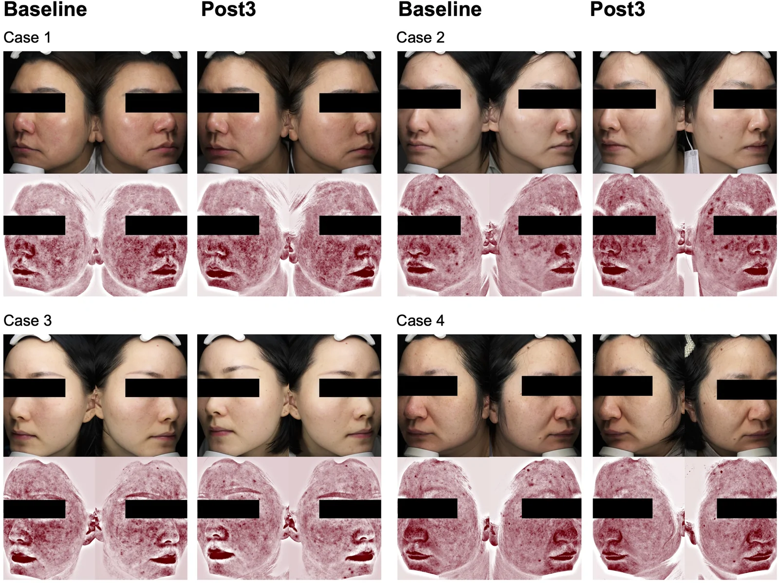
A total of 227 patients with ETR who completed three regular IPL sessions and had paired baseline and post-treatment VISIA images were included in the final analysis (Supplementary Figure 1). Representative paired clinical photographs and corresponding VISIA erythema maps before and after treatment are shown in Figure 1 . Baseline characteristics are summarized in Table 1 . The mean age was 34.1 ± 5.6 years, and 193 (85.0%) patients were female. Fitzpatrick skin type III was predominant (172/227, 75.8%), followed by type IV (55/227, 24.2%). The mean disease duration was 2.9 ± 2.2 years. The mean interval between baseline and post3 assessment was 93.4 ± 5.5 days. At baseline, the mean VISIA erythema area was 2737.55 ± 428.87 mm 2 , the mean erythema concentration was 11.55 ± 1.43%, and the median consensus CEA score was 3 (IQR, 3–4).
Table 1 Baseline Characteristics of the Enrolled Patients with Erythematotelangiectatic Rosacea
| Characteristic | Value |
|---|---|
| Age, years | |
| Mean ± SD | 34.1 ± 5.6 |
| Min–max | 25.0–43.0 |
| Sex, n (%) | |
| Male | 34 (15.0) |
| Female | 193 (85.0) |
| Fitzpatrick skin type, n (%) | |
| Type III | 172 (75.8) |
| Type IV | 55 (24.2) |
| Disease duration, years, mean ± SD | 2.9 ± 2.2 |
| Prior treatment, n (%) | |
| Oral tetracycline | 82 (36.1) |
| Topical vasoconstrictors | 33 (14.5) |
| Topical anti-inflammatory agents | 152 (67.0) |
| Interval from baseline to post3 (days), Mean ± SD | 93.4 ± 5.5 |
| Baseline erythema area (mm 2 ), Mean ± SD | 2737.55 ± 428.87 |
| Baseline erythema concentration (%), Mean ± SD | 11.55 ± 1.43 |
| Baseline consensus CEA score, Median (IQR) | 3 (3–4) |

Figure 1 Representative paired facial images before and after 3 regular IPL sessions. Representative clinical photographs and corresponding VISIA erythema maps obtained at baseline and post3 from four patients with erythematotelangiectatic rosacea, illustrating diverse erythema distributions and treatment responses.
Blinded CEA scoring demonstrated good-to-excellent inter-rater reliability at baseline and post3, with mean pairwise quadratic weighted κ values of 0.795 and 0.863, respectively (Supplementary Figure 2). Exact agreement among all three raters was observed in 195 of 227 assessments (85.9%) at baseline and 174 of 227 assessments (76.7%) at post3. Baseline ratings were predominantly distributed within CEA scores of 3 and 4, whereas post3 ratings shifted toward lower severity categories. These findings support the reproducibility of blinded CEA assessment as the clinical reference used in the present analysis.
After three regular IPL sessions, significant reductions were observed in VISIA-derived erythema parameters and consensus CEA scores. Erythema area, erythema concentration, and CEA scores all decreased significantly from baseline to post3 ( Figure 2 , all p < 0.001).
Based on the predefined responder criterion, 109 of 227 patients (48.0%) achieved a ≥2-grade reduction in CEA score and were classified as having a clinically substantial response, whereas the remaining 118 patients (52.0%) achieved a 1-grade reduction. Patients with greater CEA improvement demonstrated larger reductions in VISIA-derived erythema parameters.
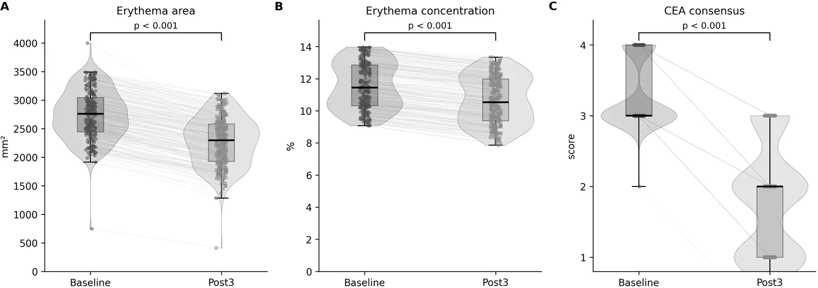
Figure 2 Changes in VISIA erythema metrics and CEA after 3 regular IPL sessions. Violin-box plots with paired trajectories show baseline and post3 values for erythema area (A), erythema concentration (B), and consensus Clinician Erythema Assessment (CEA) score (C). Wilcoxon signed-rank tests showed significant reductions in all three measures (all p < 0.001).
Changes in VISIA-derived erythema metrics were significantly associated with the magnitude of clinical improvement measured by ΔCEA ( Figure 3 ). Reduction in erythema area showed a moderate correlation with ΔCEA (Spearman ρ= 0.476, p < 0.001), whereas reduction in erythema concentration showed a weaker but significant correlation (Spearman ρ= 0.308, p < 0.001).
When patients were stratified according to clinical response, those with ΔCEA ≥2 showed significantly greater reductions in both erythema area and erythema concentration compared with those with ΔCEA =1 (both p <0.001). The stronger association observed for erythema area suggests that spatial reduction of erythematous involvement may better reflect clinician-perceived improvement than changes in erythema intensity alone.
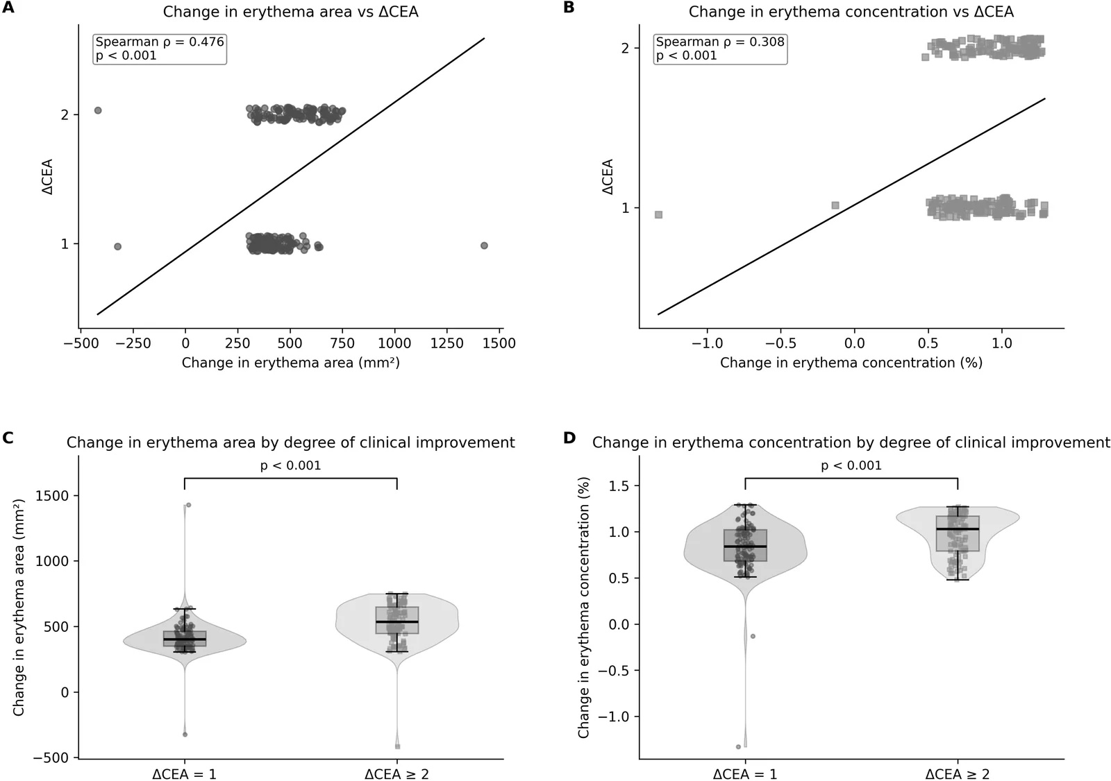
Figure 3 Relationship between VISIA-derived erythema changes and clinician-rated improvement. Scatter plots show the associations of changes in erythema area (A) and erythema concentration (B) with ΔCEA. Violin-box plots compare changes in erythema area (C) and erythema concentration (D) between patients with ΔCEA = 1 and those with ΔCEA ≥ 2.
ROC analysis was performed to identify VISIA-derived thresholds corresponding to clinically substantial improvement (ΔCEA ≥2) ( Figure 4 ). Among absolute change parameters, reduction in erythema area showed better discrimination than reduction in erythema concentration, with AUCs of 0.775 (95% CI, 0.711–0.835) and 0.678 (95% CI, 0.604–0.747), respectively.
For percentage changes, percentage reduction in erythema area demonstrated the highest discriminative performance among the evaluated VISIA parameters, with an AUC of 0.851 (95% CI, 0.797–0.901). The cohort-derived optimal cutoff was an 18.76% reduction in erythema area, yielding a sensitivity of 0.72 and specificity of 0.93. The bootstrap-derived optimal cutoffs showed a narrow distribution, with a median of 18.98% and a 95% percentile interval of 18.76%–19.68%. In comparison, percentage reduction in erythema concentration showed a lower AUC of 0.715 (95% CI, 0.645–0.782).
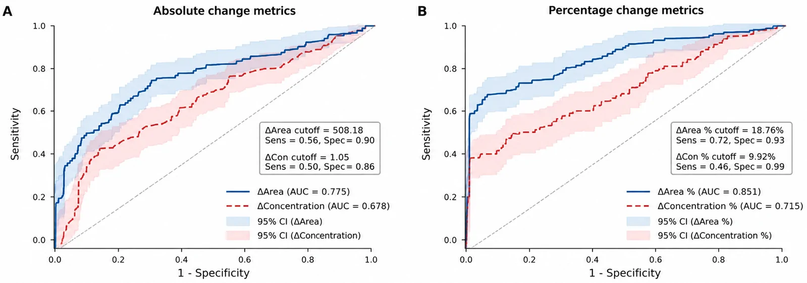
Figure 4 Receiver operating characteristic (ROC) analysis of VISIA-derived metrics for identifying clinically substantial improvement (ΔCEA ≥2). (A) Absolute changes in erythema area and erythema concentration. (B) Percentage changes in erythema area and erythema concentration. AUCs (95% CIs) were 0.775 (0.711–0.835) for absolute erythema area reduction, 0.678 (0.604–0.747) for absolute erythema concentration reduction, 0.851 (0.797–0.901) for percentage erythema area reduction, and 0.715 (0.645–0.782) for percentage erythema concentration reduction. Optimal cutoffs were determined using the Youden index. Shaded areas represent 95% confidence intervals.
In this real-world cohort study of 227 patients with ETR, we evaluated the relationship between objective VISIA-derived erythema metrics and blinded clinician assessment after regular IPL therapy. Three major findings were observed. First, blinded CEA demonstrated good-to-excellent inter-rater reliability, supporting its reproducibility as a clinician-based reference for erythema assessment. Second, VISIA-derived erythema parameters showed significant responsiveness after IPL treatment and correlated with the magnitude of clinical improvement assessed by ΔCEA. Third, ROC analysis identified percentage reduction in erythema area as the most discriminative VISIA-derived parameter for identifying patients with a ≥2-grade reduction in CEA score, with an optimal cutoff of 18.76%. This extends our previous work on the cumulative multidimensional effects of long-term IPL treatment by shifting the focus from treatment efficacy and predictors of overall aesthetic response to the evaluation and clinical interpretability of objective erythema measurement in ETR. 19
Assessment of persistent erythema remains challenging in ETR because diffuse facial redness is difficult to quantify using conventional clinical evaluation alone. The CEA grading scale is widely used because of its simplicity and clinical interpretability; however, ordinal grading may be influenced by observer experience, particularly when distinguishing adjacent severity categories. 11 In the present study, blinded CEA scoring showed substantial agreement among dermatologists, with quadratic weighted κ values of 0.795 at baseline and 0.863 after treatment. These findings support the reliability of blinded CEA assessment when standardized image review and consensus evaluation are applied. Therefore, CEA may serve as a clinically meaningful anchor for evaluating objective imaging-based measures.
Digital imaging technologies may provide complementary approaches for improving objectivity in erythema assessment. 16–18 In this study, both VISIA erythema area and erythema concentration decreased significantly after IPL therapy, and their changes were associated with clinician-rated improvement. Notably, erythema area demonstrated a stronger correlation with ΔCEA than erythema concentration and achieved superior performance in ROC analysis. This finding suggests that the spatial extent of erythematous involvement may better reflect overall clinical improvement than changes in erythema intensity alone. 16 , 17 From a practical perspective, reduction in erythema area may capture the global visual improvement recognized by clinicians when assessing facial erythema severity.
An important contribution of this study is the identification of a candidate VISIA-based threshold linked to clinician-rated improvement. Rather than assessing only statistical changes in erythema parameters, we evaluated whether quantitative imaging changes could distinguish patients with different degrees of clinical response. A percentage reduction of 18.76% in VISIA erythema area provided the best discrimination for a ≥2-grade CEA reduction. This threshold should be interpreted as a responder criterion derived from this cohort rather than a validated minimal clinically important difference, because patient-reported outcomes and external validation were not available. 22 , 23 Nevertheless, it provides a candidate reference for future prospective validation studies.
The integration of clinician-based assessment and objective imaging may improve the evaluation of vascular-directed therapies in rosacea. 16–18 While clinical scores remain essential because they reflect expert interpretation of disease severity, imaging-derived metrics can provide continuous quantitative information that may enhance longitudinal monitoring. 16–18 The present findings suggest that VISIA-derived erythema measurements, particularly area-based parameters, may serve as objective adjuncts rather than replacements for clinician assessment.
Several limitations should be acknowledged. First, this was a retrospective single-center study, and the findings require validation in prospective multicenter cohorts. Second, although treatment and assessment intervals were relatively standardized, IPL parameters were individualized according to clinical characteristics, reflecting routine practice but introducing potential heterogeneity. Third, patient-reported outcomes were unavailable; therefore, the relationship between objective erythema reduction, clinician-rated improvement, and patient-perceived benefit could not be evaluated. Future studies incorporating quality-of-life measures and patient satisfaction assessments are needed. 24 , 25 Finally, VISIA measurements depend on standardized image acquisition and proprietary algorithms, which may limit direct comparability across different imaging platforms.
In conclusion, in this retrospective single-center cohort, VISIA-derived erythema metrics, particularly erythema area reduction, correlated with blinded clinician-assessed improvement after IPL therapy in ETR and may serve as objective adjuncts for treatment-response monitoring. The cohort-derived 18.76% threshold should be considered exploratory rather than a validated minimal clinically important difference and requires external validation, ideally in prospective multicenter studies incorporating patient-reported outcomes.
声明:中文精读 · 仅供学术参考。内容来自公开文献检索,不代表本人观点,不构成诊疗建议。 医疗美容需在正规医疗机构由执业医师实施。
基于相同主题推荐 · 继续深挖
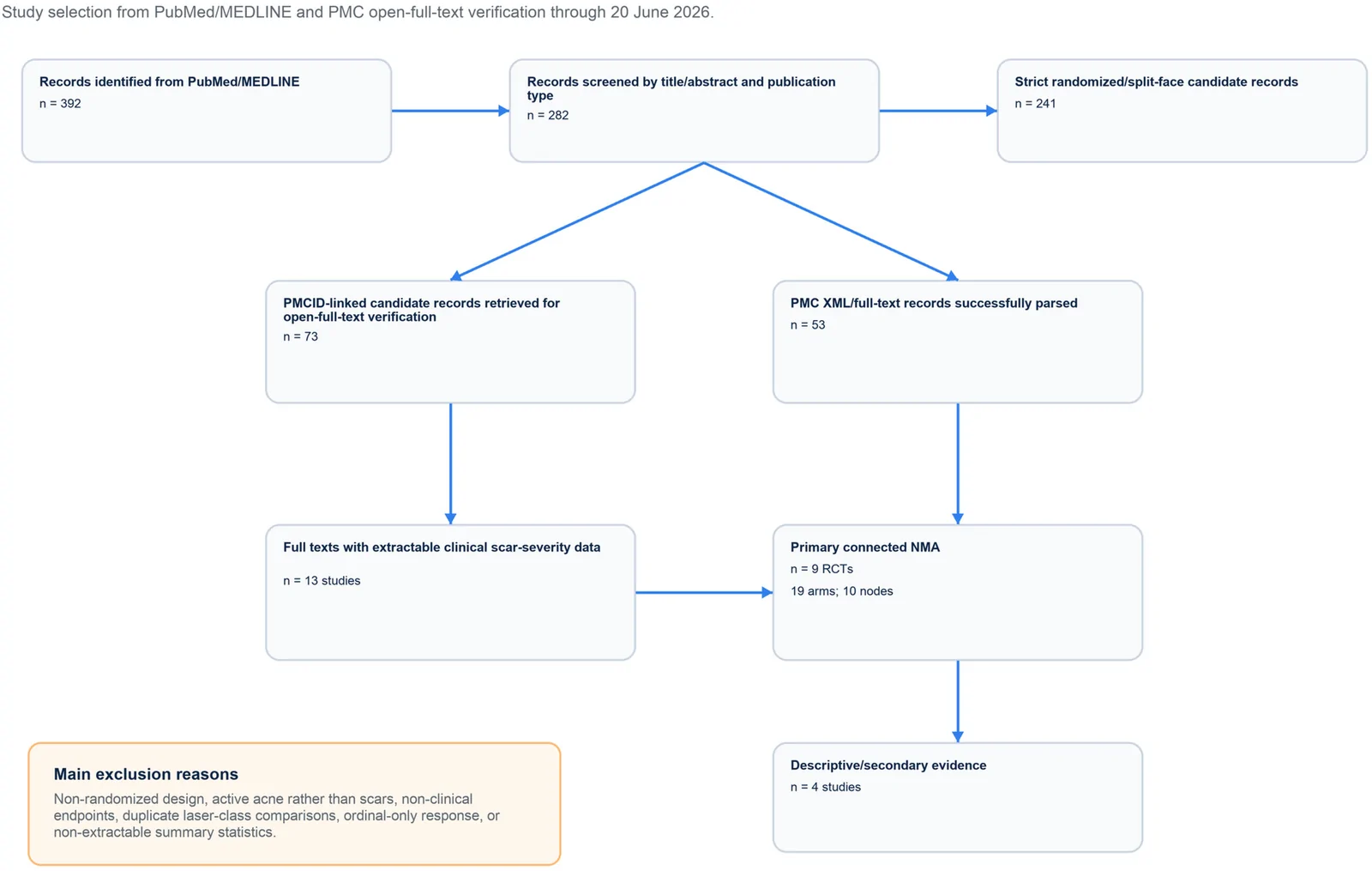
PROSPERO注册的系统综述,纳入9项随机/分脸随机试验、19个治疗臂,构成稀疏连接网络并采用频率学随机效应网络荟萃分析。
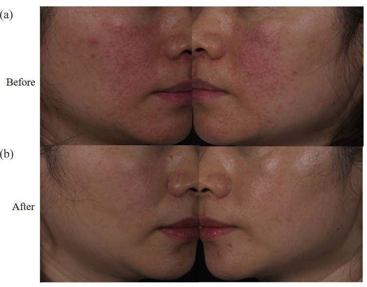
回顾性纳入63例红斑毛细血管扩张型玫瑰痤疮(ETR)中国患者,均采用555–950 nm与530–750 nm双滤光片窄谱IPL治疗。
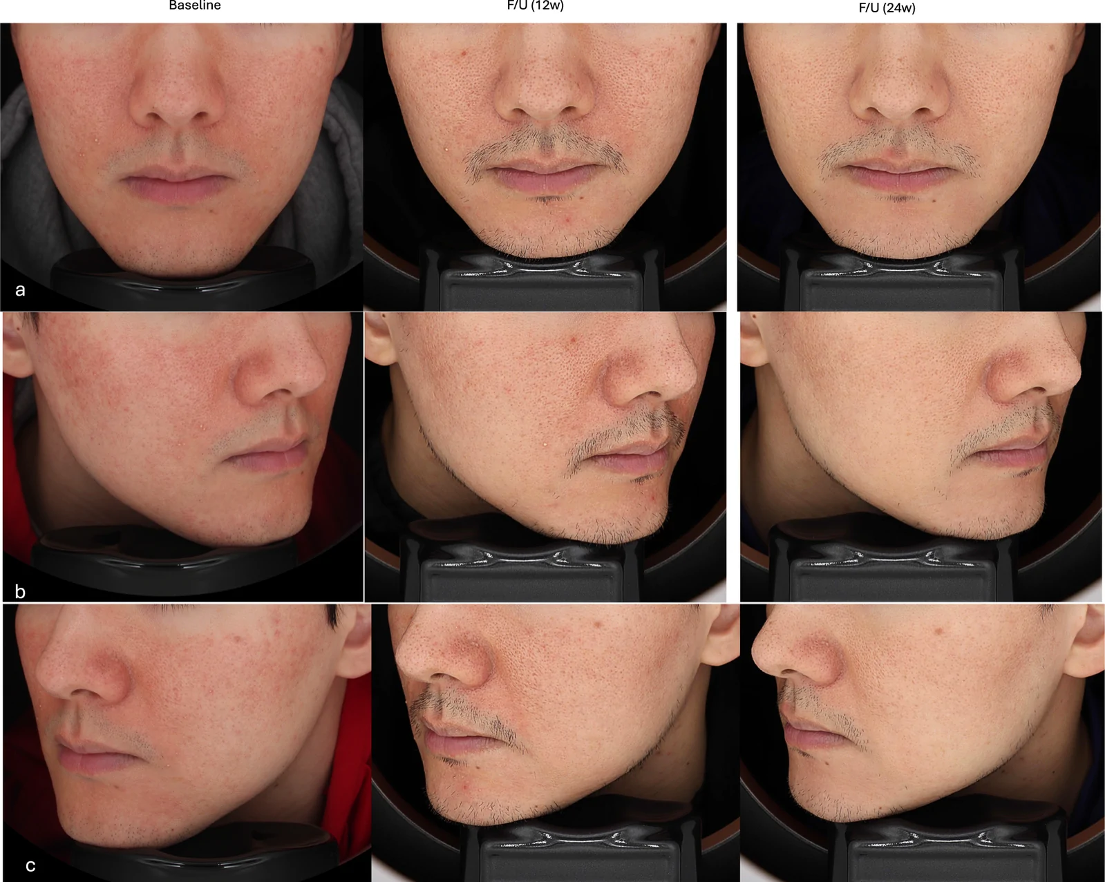
真实世界回顾性队列纳入50例临床诊断玫瑰痤疮成人,80%为女性、中位年龄33岁,多数(76%)为Fitzpatrick III型肤色。