有限剥离保留韧带面部提升术:早期面部老化的术式与技术
2026年10月16日25 min read
Aesthetic Surgery Journal. Open Forum

结论与实践要点
以下为基于原文的独立补充解读,不替代原文,具体临床决策请结合原文与自身评估。
BackgroundEarly facial aging is characterized by midfacial descent, loss of mandibular definition, and attenuation of retaining ligaments. Although superficial musculoaponeurotic system (SMAS)-based and deep-plane facelifts remain the gold standard, many patients seek less invasive procedures with shorter recovery.
ObjectivesThe aim of this study was to describe a limited-dissection ligament-preserving facelift technique and evaluate its safety and clinical outcomes in patients with early facial aging.
MethodsA retrospective case series of 100 patients treated between June 2021 and January 2025 was performed. All procedures were carried out under local anesthesia using limited dissection and superoposterior suspension of the SMAS–ligament complex and platysma. Outcomes included complications, revision rates, recovery time, and patient satisfaction at 1 year.
ResultsMean patient age was 43.4 years (range, 34-52 years). All procedures were performed under local anesthesia without sedation. Recovery was rapid, with 96% of patients resuming normal activities within 7 days. Transient hypoesthesia occurred in 22% of patients and resolved within 8 weeks. No hematoma, infection, permanent nerve injury, or wound dehiscence was observed. Temporal alopecia occurred in 14% of patients and resolved spontaneously in most cases. Minor under-correction was observed in 6%, with an overall revision rate of 9%. At 1 year, 82% of patients reported being satisfied or very satisfied.
ConclusionsLimited-dissection ligament-preserving facelift appears to be a safe and effective minimally invasive option for selected patients with early facial aging. It provides natural results and rapid recovery and may represent an intermediate approach between nonsurgical treatments and traditional facelift procedures.
Level of Evidence: 4 (Therapeutic)For image description, please refer to the figure legend and surrounding text.
Facial aging is a progressive and multifactorial process that affects the skin, soft tissues, and underlying skeletal support, beginning as early as the third to fourth decade of life. 1 , 2 Early manifestations include midfacial ptosis, deepening of the nasolabial folds, and loss of mandibular definition. 3 These changes are largely attributed to the descent of the superficial musculoaponeurotic system (SMAS), attenuation and elongation of retaining ligaments, and progressive loss of dermal elasticity. 4 , 5
Recent anatomical studies suggest that facial aging may not be solely explained by gravitational descent or increased tissue mobility. Several authors have proposed that attenuation, elongation, and loss of functional support within the retaining ligament system may contribute to early soft-tissue descent by reducing structural tension across the superficial fascial-ligamentous network. 6 Although the precise contribution of retaining ligament laxity to facial aging remains incompletely understood, these observations support the hypothesis that restoration of support within the SMAS–ligament complex may contribute to facial rejuvenation in selected patients without requiring extensive deep-plane mobilization. Further anatomical and clinical studies are needed to better define the role of this mechanism.
Facial rejuvenation surgery has evolved substantially following the characterization of the SMAS and the functional role of facial retaining ligaments. Traditional SMAS-based rhytidectomy techniques and deep-plane facelifts remain well-established procedures, providing predictable and long-lasting outcomes. 7-10 However, a significant proportion of patients with early signs of aging are reluctant to undergo extensive surgical procedures under general anesthesia due to prolonged recovery, higher morbidity, and associated risks. 11 Consequently, both nonsurgical and minimally invasive surgical approaches have emerged, aiming to achieve early correction of facial descent while preserving natural anatomical relationships and minimizing downtime. 12 , 13
This study describes a minimally invasive facelift technique designed to achieve vertical midfacial elevation and SMAS-platysma suspension through 4 limited incisions, performed under local anesthesia without hospitalization. The objectives are to detail the surgical technique, define patient selection criteria, evaluate clinical outcomes, and position this approach within the contemporary facial rejuvenation paradigm.
A retrospective case series was conducted, including 100 patients who underwent limited-dissection ligament-preserving facelifts between June 2021 and January 2025 at a private clinic. All procedures were performed in accordance with the ethical principles of the Declaration of Helsinki and local clinical practice guidelines. Written informed consent was obtained from all patients for both the surgical procedure and the use of anonymized clinical data and photographic documentation for academic purposes. Patients were selected based on the presence of early signs of facial aging, including midfacial descent, loss of mandibular definition, and nasolabial volume redistribution, in the absence of significant cervical laxity or platysmal banding. Exclusion criteria included advanced facial aging requiring more extensive surgical correction, marked submental adiposity, and significant fibrosis related to previous procedures. Clinical outcomes were assessed, including photographic evaluation, complication and revision rates, and patient-reported outcomes obtained through a standardized, nonvalidated satisfaction survey administered at 1-year follow-up.
A subgroup of 20 patients with standardized preoperative and 12-month postoperative photographs underwent blinded evaluation by 3 independent facial plastic surgeons using a 4-point aesthetic improvement scale (0 = no improvement; 1 = mild improvement; 2 = moderate improvement; 3 = significant improvement).
This physician-rated scale was developed for the purpose of this study and has not been formally validated. Scores were assigned according to the degree of improvement in mandibular definition, jawline contour, and midfacial support. No validated reference atlas was available.
Reviewers were provided with paired preoperative and postoperative images without access to patient identity or satisfaction data.
Standardized photographic documentation suitable for physician assessment was available only in a subgroup of 20 patients. Because of the retrospective nature of the study, photographic acquisition protocols were not uniform across the entire cohort. Therefore, physician-rated photographic assessment was limited to patients with complete standardized image sets.
Descriptive statistical analysis was performed to evaluate demographic characteristics, recovery time, complication rates, and overall patient satisfaction. All patients completed the 1-year follow-up assessment.
Facial soft tissues can be conceptualized as a continuous tension-bearing system composed of the skin, SMAS, ligamentous septa, and deep fascia. Classical anatomical studies have demonstrated that the SMAS represents a specialized layer of the superficial fascia, linking the mimetic musculature to the dermis, while retaining ligaments integrate this system with the underlying skeletal framework. 4 , 5
Age-related elongation and attenuation of retaining ligaments may contribute to reduced structural support within the superficial fascial-ligamentous network. From this perspective, early facial aging may primarily reflect a loss of tension within the superficial fascial-ligamentous network rather than a simple increase in tissue laxity or mobility.
Retaining ligaments originate from deep fascial and periosteal attachments, traverse the SMAS layer, and insert into the dermis through cutaneous extensions. In the present technique, selective release is performed at the level of the cutaneous ligamentous attachments while preserving the deeper ligament–SMAS continuity. The remaining SMAS–ligament complex is then utilized as a structurally reinforced suspension point for tissue repositioning. This approach may provide stronger and more anatomically stable fixation compared with suspension performed through the SMAS alone.
Identification of cutaneous ligamentous structures during limited dissection allows the underlying SMAS–ligament complex to be utilized as a stable anchoring platform for tissue repositioning. The surgical approach is therefore based on superoposterior suspension of the SMAS–ligament complex, while preserving the majority of deep facial-retaining ligaments, thereby maintaining structural integrity and minimizing surgical trauma.
The surgical technique is demonstrated in Video 1, showing dissection and SMAS suspension in the facial region, and Video 2, demonstrating cervical dissection and platysma suspension.
Two symmetrical temporal incisions are designed in an oblique orientation, each measuring ∼2 to 3 cm in length and positioned perpendicular to the planned vector of traction. The incisions are placed within the trichophytic zone of the temporal scalp, at least 1 cm posterior to the anterior hairline, and oriented parallel to the direction of hair follicles to optimize camouflage and achieve near-invisible scarring. This approach provides optimal access to the mobile SMAS below the zygomatic arch and the malar fat pad while maintaining concealment within the hair-bearing scalp. Two additional incisions (2-3 cm) are placed in the postauricular sulcus, carefully positioned posterior to the auricular border and never extending anterior to the earlobe. These incisions allow access for suspension of the lateral border of the platysma while preserving an inconspicuous scar in aesthetically sensitive areas.
The procedure is performed under local anesthesia without sedation. A solution of 2% lidocaine with epinephrine (1:100,000) is infiltrated into the planned incision sites and along the anticipated dissection planes, extending ∼3 to 4 cm from the incision margins. This provides effective analgesia, vasoconstriction, and hydrodissection, facilitating atraumatic tissue separation.
Following skin incision with a No. 15 scalpel blade, blunt dissection is performed through the subcutaneous tissue to reach the SMAS in the facial region and the superficial cervical fascia with the platysma in the cervical region. Cutaneous retaining ligaments are selectively identified and released bilaterally to allow mobilization, while preserving the integrity of the SMAS–ligament complex.
Dissection is deliberately limited and targeted. In the facial region, it focuses on mobilization of the SMAS and malar fat pad; in the cervical region, it addresses the lateral border of the platysma ( Figure 1 ). The extent of ligament release was determined intraoperatively according to tissue mobility and the amount of superoposterior repositioning required to achieve adequate correction, typically requiring 3 to 5 cm of dissection from the incision site to reach the mobile SMAS and platysma.
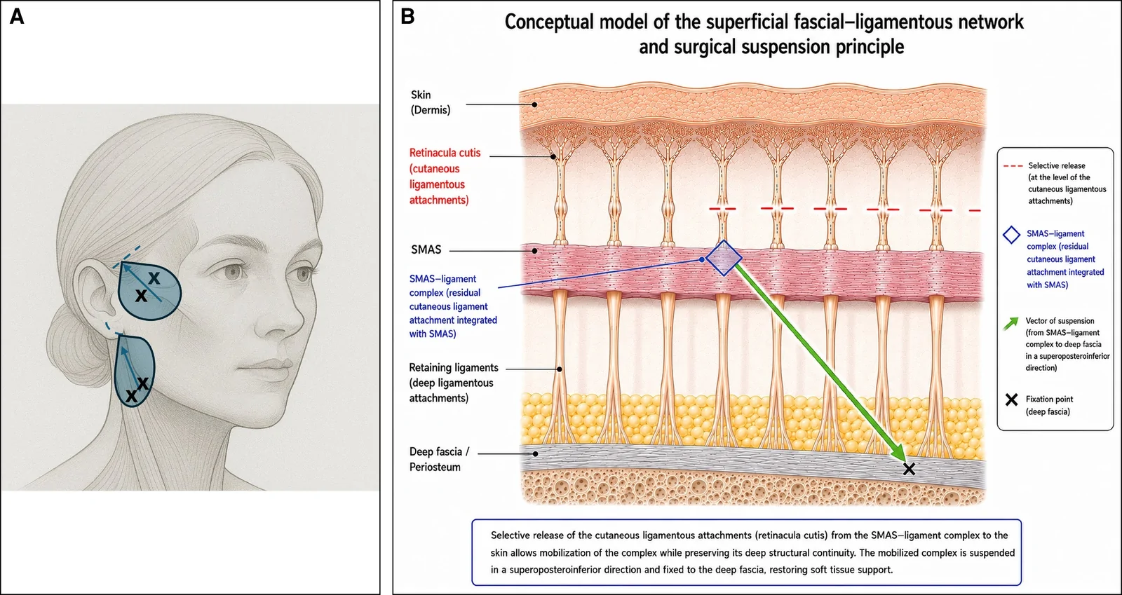
Figure 1. (A) Schematic representation of the surgical technique, illustrating incision placement (dotted lines), areas of dissection (blue zones), and fixation points (X). (B) Diagram of the facial retaining ligaments, demonstrating selective release of the cutaneous component (retinacula cutis) while preserving the SMAS–ligament complex (rhombus) as a stable anchoring structure. This conceptual illustration was created with the assistance of ChatGPT (OpenAI, San Francisco, CA) and subsequently reviewed and edited by the authors.
Once adequate mobilization has been achieved, 2 permanent 2-0 polypropylene sutures are placed to suspend the SMAS–ligament complex to the deep temporal fascia. The suspension vector is oriented in a predominantly superoposterior direction ( Figure 2 ).
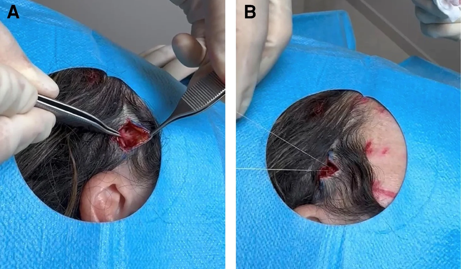
Figure 2. (A) Identification and release of the cutaneous ligaments in the facial region. (B) Suspension of the SMAS–ligament complex in the facial region.Two additional permanent 2-0 polypropylene sutures are used to suspend the lateral border of the platysma to the deep cervical fascia. This maneuver enhances the definition of the mandibular border and improves the cervicomental angle while maintaining a minimally invasive approach ( Figure 3 ).
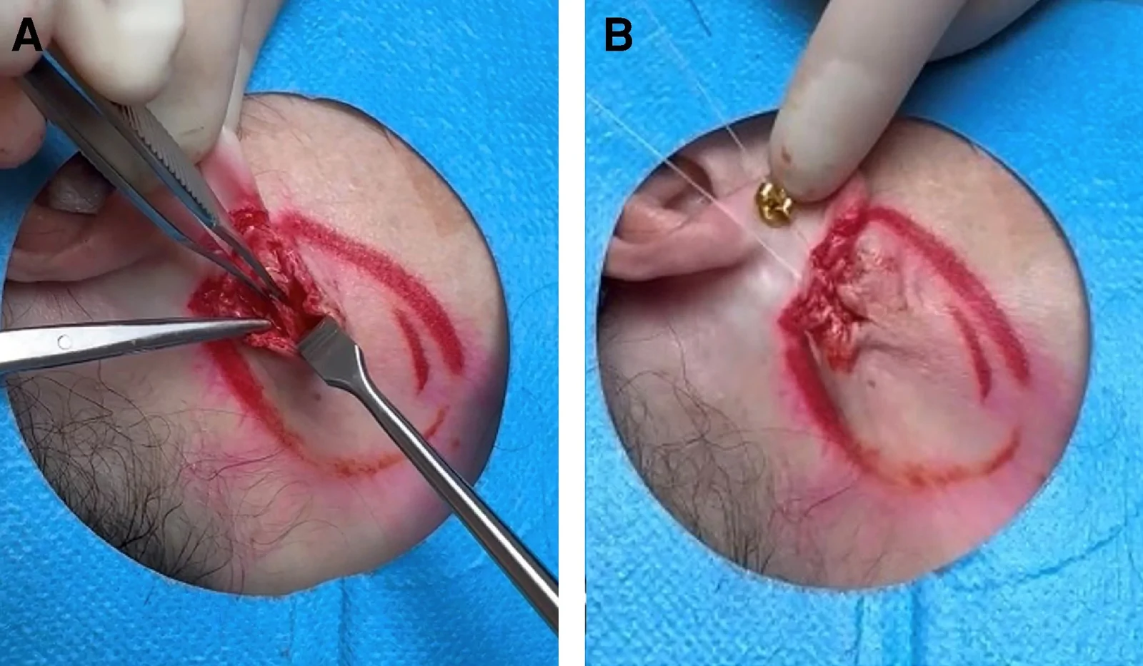
Figure 3. (A) Identification and release of the cutaneous ligaments in the cervical region. (B) Suspension of the platysma (right side) in the cervical region.Permanent fixation was intentionally selected because the technique relies on limited dissection and suture-based suspension rather than wide SMAS flap elevation. Depending on tissue laxity, ∼2 cm of SMAS advancement and folding were achieved during suspension.
Following fixation, residual skin redundancy is carefully assessed, and when indicated, a conservative elliptical skin excision (∼1 - 2 cm in width) is performed. Wound closure is carried out in 2 layers, with the subcutaneous tissue approximated using 4-0 absorbable sutures and the dermis closed with 5-0 interrupted polypropylene sutures. No surgical drains are required due to the limited extent of dissection and minimal bleeding risk. A light compressive dressing is applied at the conclusion of the procedure and removed after 24 h.
Patients are discharged on the same day of surgery. Postoperative instructions include application of cold compresses during the first 24 h and maintenance of head elevation for 48 h. Sutures are removed on postoperative Day 7.
Between June 2021 and January 2025, a total of 100 patients underwent the minimally invasive facelift procedure. The cohort consisted of carefully selected individuals presenting with early signs of facial aging, including midfacial descent, loss of mandibular definition, and nasolabial volume redistribution, without significant cervical skin laxity or platysmal banding. No concomitant surgical procedures were performed at the time of the facelift, allowing isolated evaluation of the technique ( Figures 4 , 5 ). The mean patient age was 43.4 years (range, 34-52 years), and all procedures were performed under local anesthesia without sedation (100%). The procedure was well tolerated in all cases, with an uneventful postoperative course in the majority of patients. Mild edema and ecchymosis were common but resolved spontaneously within 5 to 7 days. A total of 96% of patients resumed work or normal social activities within 7 days, despite mild residual bruising in some cases. No surgical drains were required, and no patient required narcotic analgesia beyond 48 h postoperatively. At 1-year follow-up, patient-reported outcomes demonstrated high levels of satisfaction, with 82% of patients reporting being satisfied or very satisfied with the aesthetic results ( Table 1 ), based on a standardized survey distributed through email to all patients at 1 year postoperatively.
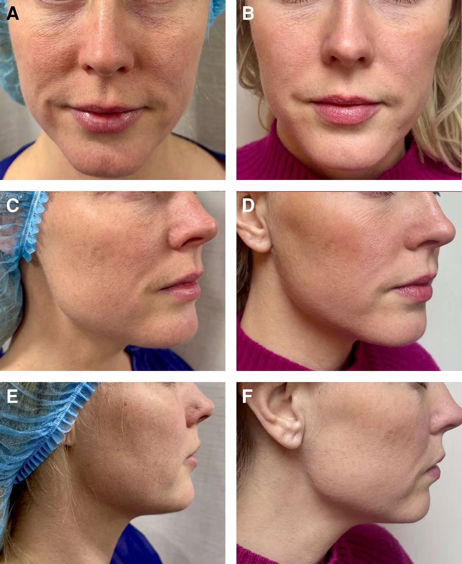
Figure 4. (A) Preoperative frontal view of a 41-year-old female patient. (B) Postoperative frontal view at 12 months following limited-dissection ligament-preserving facelift. (C) Preoperative right 3-quarter view. (D) Postoperative right 3-quarter view at 12 months. (E) Preoperative right lateral view. (F) Postoperative right lateral view at 12 months.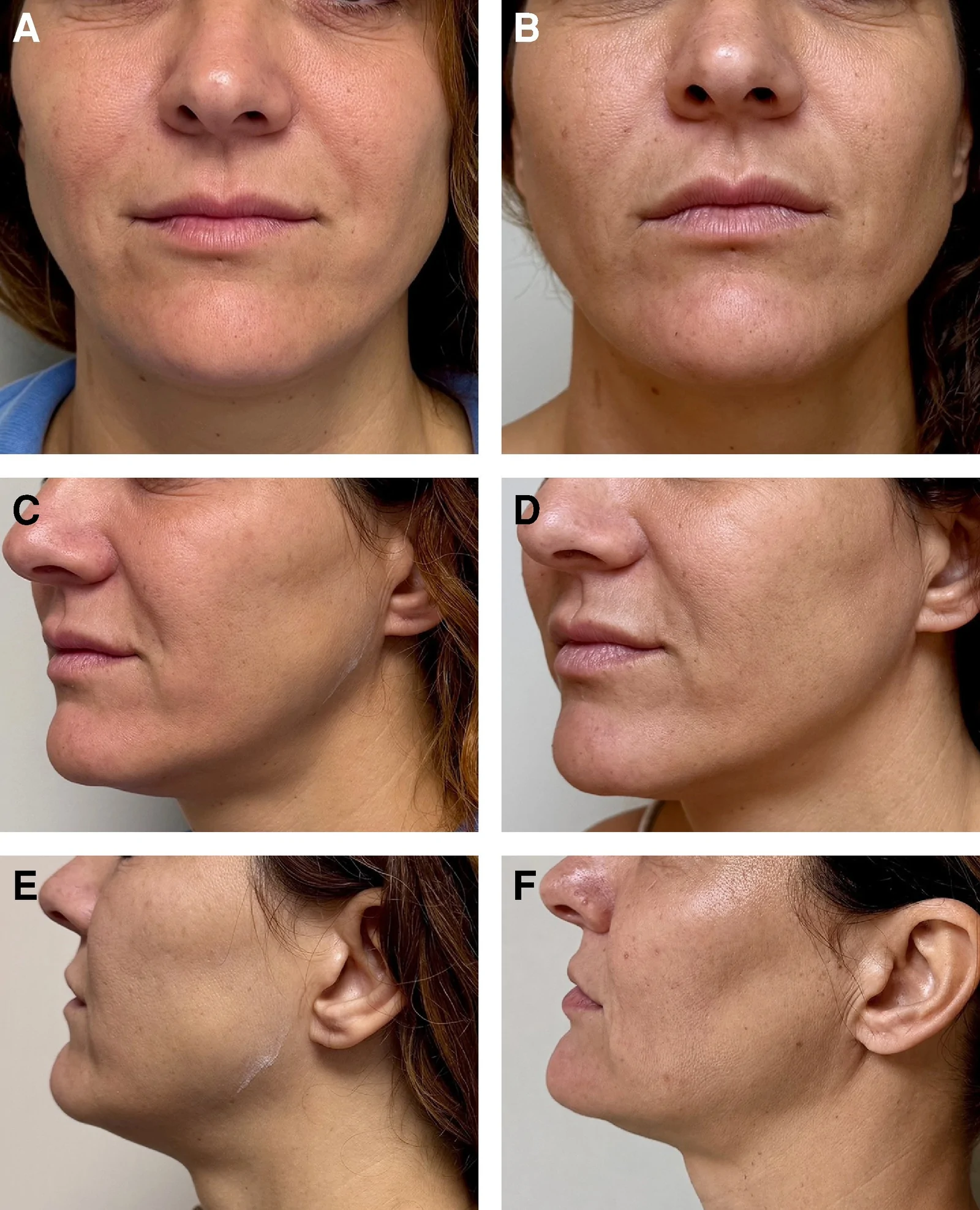
Figure 5. (A) Preoperative frontal view of a 45-year-old female patient. (B) Postoperative frontal view at 12 months following limited-dissection ligament-preserving facelift. (C) Preoperative left 3-quarter view. (D) Postoperative left 3-quarter view at 12 months. (E) Preoperative left lateral view. (F) Postoperative left lateral view at 12 months.Table 1. Patient Satisfaction at 1-year Follow-up
| Satisfaction level | No. of patients ( n = 100) |
|---|---|
| Very satisfied | 31 |
| Satisfied | 51 |
| Neutral | 11 |
| Dissatisfied | 6 |
| Very dissatisfied | 1 |
Blinded photographic evaluation of the subgroup demonstrated predominantly moderate or significant improvement in mandibular definition and midfacial support ( Table 2 ).
Table 2. Photographic Evaluation
| Aesthetic improvement score | No. of patients (%) |
|---|---|
| Significant improvement (3) | 8 (40) |
| Moderate improvement (2) | 9 (45) |
| Mild improvement (1) | 3 (15) |
| No improvement (0) | 0 (0) |
Transient hypoesthesia occurred in 22% of patients ( n = 22), primarily in the temporal and preauricular regions, with complete resolution in all cases within 8 weeks. No cases of permanent nerve injury, hematoma, infection, or wound dehiscence were observed. Temporal alopecia at the incision site was noted in 14% of patients ( n = 14), resolving spontaneously within 3 months in 11 cases, whereas 3 patients required minor surgical revision. Minor under-correction was observed in 6% of patients ( n = 6), all of whom underwent secondary revision procedures. In addition, 3 patients required revision for persistent temporal alopecia. The overall revision rate was therefore 9% ( n = 9).
Facial rejuvenation techniques have evolved considerably in recent years, largely driven by increasing patient demand for effective outcomes with reduced downtime and lower procedural risk. Although deep-plane and traditional SMAS-based facelifts remain the gold standard for addressing advanced facial aging, a growing proportion of patients in their fourth decade, despite being suitable candidates, are reluctant to undergo extensive surgical procedures under general anesthesia. This trend has created a clear clinical need for minimally invasive yet structurally effective alternatives.
The present technique reflects a conceptual shift in the understanding of early facial aging. In many patients in their 40s, one possible contributing mechanism may be progressive attenuation of support within the superficial fascial-ligamentous network. Although deep-plane approaches remain highly effective for advanced aging, minimally invasive facelift techniques may serve as an intermediate solution, bridging the gap between nonsurgical treatments and conventional facelifts.
Minimally invasive facelift itself is not a novel concept, having been described for more than 5 decades. 14 , 15 Historically, both patients and surgeons have sought facial rejuvenation techniques that minimize incisions and recovery time. Over the past decade, nonsurgical modalities have gained widespread popularity; however, in some cases, they have overpromised and failed to deliver consistent or durable outcomes. Increasingly, patients report dissatisfaction with thread-lifting procedures and excessive use of hyaluronic acid fillers. Thread lifts, in particular, have been associated with partial relapse within 6 to 12 months and complication rates of up to 17%. 16 , 17 Similarly, overcorrection with fillers may distort natural facial proportions, leading to an unnatural and volumized appearance of the midface.
In clinical practice, this has translated into a growing population of younger patients seeking subtle yet meaningful improvements without the burden of prolonged recovery or invasive surgery. Contemporary aesthetic trends further support this shift, with increasing interest in concepts such as the “micro lift” and “weekend facelift,” which emphasize rapid recovery and natural results. In this context, limited-dissection ligament-preserving facelift represents a reproducible and structurally sound approach that addresses early tissue descent while maintaining a minimally invasive profile.
The technique described in this study is based on SMAS and platysma suspension through 4 small, strategically concealed incisions.
Unlike thread-lifting procedures, which rely on percutaneous suspension without direct visualization of the deeper soft-tissue support structures, the present technique allows direct limited-access identification and fixation of the SMAS–ligament complex and lateral platysma. The clinical role of this procedure is not to replace conventional SMAS or deep-plane facelift surgery but rather to fill the therapeutic gap between nonsurgical suspension techniques and more extensive facelift procedures.
In contrast to other short-scar or minimally invasive facelift techniques, such as minimal access cranial suspension lift, S-lift, vertical lift, or micro-facelift, this approach avoids preauricular or sideburn incisions and limits the extent of dissection. 18-23 Although transcutaneous facelift techniques have also been described, direct subcutaneous dissection in the present method allows improved visualization and more precise engagement of the SMAS–ligament complex. 24 Additionally, controlled and minimal skin excision reduces the risk of unnatural skin folding, which may occur with other limited-access techniques. It should be acknowledged, however, that there is currently no standardized nomenclature for minimally invasive facelift procedures, and the development of a unified classification system would be beneficial for both clinical practice and research.
Fixation represents a critical determinant of long-term outcomes. Suspension of the SMAS–ligament complex and platysma may contribute to improvement of soft-tissue support, rather than relying solely on skin redraping. This approach may contribute to improved durability while maintaining a favorable safety profile, with minimal risk of nerve injury due to the limited depth of dissection.
Vector orientation is another key factor influencing aesthetic outcomes. The superoposterior vector employed in this technique contributes to restoration of malar projection and mandibular definition while minimizing distortion of natural facial contours. The predominantly vertical component of the lift also reduces skin redundancy, thereby limiting the need for larger skin excisions and longer incisions.
An additional advantage of this technique is its feasibility under local anesthesia in an outpatient setting. The relatively short operative time (typically <1 h) and minimal intraoperative discomfort eliminate the need for sedation in most cases, further contributing to reduced procedural risk and faster recovery. In selected patients with mild submental adiposity, this procedure may be combined with conservative submental liposuction. However, no concomitant liposuction was performed in the present series.
Despite these advantages, several limitations must be recognized. This technique is not intended to replace deep-plane or traditional facelifts, particularly in patients with advanced facial aging. Improvements in the cervicomental angle and mandibular contour may be limited, especially in individuals with significant platysmal banding or submental adiposity. Skin excision is intentionally conservative (1-2 cm), which may restrict correction in patients with more advanced laxity. Additionally, the longevity of results is expected to be shorter than that achieved with more extensive surgical techniques and may vary among patients. Careful patient selection is therefore essential to ensure appropriate indications and to avoid unrealistic expectations. Finally, patients with significant facial fibrosis resulting from previous interventions may represent a relative contraindication due to impaired tissue mobility.
The present study has several limitations. Its retrospective design, lack of control group, and reliance on a nonvalidated patient satisfaction survey limit the strength of conclusions. Objective anthropometric or 3D image measurements and validated patient-reported outcome instruments were not routinely available due to the retrospective nature of the study. Further prospective comparative studies with standardized outcome measures are warranted.
The proportion of neutral or dissatisfied patients likely reflects both the intentionally conservative nature of the technique and variability in patient expectations. Patients seeking correction comparable to a conventional SMAS or deep-plane facelift may perceive the improvement as insufficient despite objective or surgeon-perceived improvement. This finding emphasizes the importance of careful patient selection and preoperative counseling regarding the limited magnitude of correction achievable with this approach.
The 12-month follow-up should be interpreted as an early outcome assessment and does not establish long-term durability. Because this technique relies on limited dissection and permanent suture fixation rather than extensive SMAS flap elevation, longer follow-up is required to determine the stability of the aesthetic correction over time.
Limited-dissection ligament-preserving facelift represents a contemporary, minimally invasive alternative for patients in their fourth decade who present with early signs of facial aging but are reluctant to undergo extensive surgical procedures under general anesthesia and prolonged recovery. Limited-dissection ligament-preserving facelift provides a safe and effective approach in appropriately selected patients, restoring structural support and maintaining natural facial expression while avoiding aggressive surgical intervention. Careful patient selection is essential to ensure realistic expectations. This technique does not replace traditional SMAS-based or deep-plane facelifts but, in selected cases, may delay the need for more invasive procedures.
声明:中文精读 · 仅供学术参考。内容来自公开文献检索,不代表本人观点,不构成诊疗建议。 医疗美容需在正规医疗机构由执业医师实施。
基于相同主题推荐 · 继续深挖
有限剥离保留韧带面部提升术为针对早期面部老化的微创术式,回顾性病例系列纳入 2021–2025 年 100 例患者,平均 43.4 岁(范围 34–52),全部于局麻(无镇静)下完
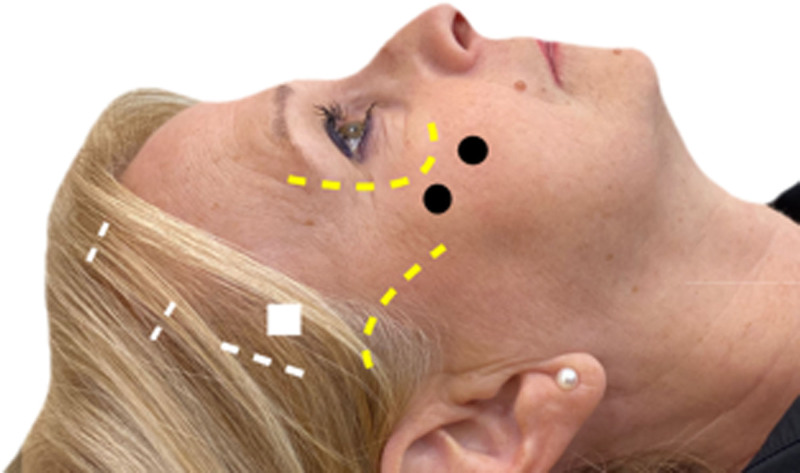
研究背景:除皱术从单层皮瓣演进到针对 SMAS(表浅肌筋膜系统)等深层结构,效果更自然持久;但经耳前大切口仍有面神经损伤与皮瓣坏死风险。本研究评估两种内镜入路(含深部 SMAS 提
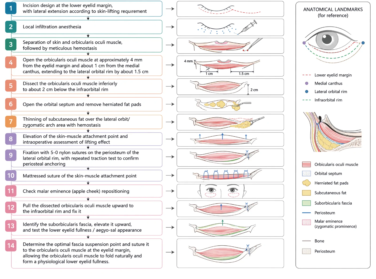
V3中面部提升经睫毛下切口,沿内眦线、外眦线、颧脂肪垫下界三个向量悬吊,可同时改善中面部下垂、下睑袋及睑颊轮廓不连续。