3D打印导板与压电颏成形术的精度分析
2026年10月21日25 min read
Plastic and Reconstructive Surgery Global Open
结论与实践要点
对需颏成形(常作为正颌手术补充或单独美学修正)者,采用基于术前CT的虚拟手术规划(VSP)与压电手术,并以3D打印生物相容树脂(BioMed Clear Resin)制作切割与复位导板,意在提升截骨与骨段复位的精度、可重复性与可预测性,减少术者依赖误差及围术期并发症。
本研究为单中心小样本(12例,11例为牙颌畸形、1例美学)、无对照组、缺乏随访,且导板形态未标准化。形态测量仅显示规划与术后3D模型的几何平均偏差(平移1.91mm、旋转右1.38/左1.30mm、颅尾右1.39/左1.23mm),代表精度而非临床疗效;成本较外协降低约€400-€800/例。结论(简单、准确、安全)需更大样本与对照研究验证,外推宜谨慎。
Background:We aimed to describe a protocol and related surgical treatment for mentoplasty using virtual surgical planning (VSP) and piezosurgery.
Methods:Twelve patients requiring mentoplasty underwent VSP based on preoperative computed tomography. Virtual osteotomy was defined and the bone fragment was repositioned. Cutting and repositioning guides were exported as STL files and created using a 3-dimensional (3D) printer with biocompatible material. Plates were preblended on the printed model. Postoperatively, morphometric analysis was performed by comparing the planned and postoperative 3D models: distance between the apex of the cusp of 43 and 33 and lower edge of the chin; distance between the lower edge of the right and left mental foramen and osteotomy; and distance between the mandibular lingual fossa and pogonion.
Results:Six cutting guides were made composite and six were monobloc. The repositioning guides were all made monobloc. No adverse intraoperative events were observed. Postoperative transient hypoesthesia was reported in 1 case. The average distance between the planned and postoperative 3D models of the chin was recorded in translation (1.91 mm), rotation (right, 1.38 mm; left, 1.30 mm), and cranial-caudal repositioning (right, 1.39 mm; left, 1.23 mm). VSP and 3D printing have improved surgical precision and reproducibility; they help the surgeon decide the extent of chin movement and the most suitable type of surgical guide in advance.
Conclusions:Mentoplasty using 3D-printed guides and piezosurgery is simple, accurate, and safe. The costs of guide production are reduced using 3D printers by approximately €400–€800 per case compared with industrial printers.
Question: What is an application of virtual surgical planning and piezosurgery in mentoplasty?
Findings: Our findings demonstrated that mentoplasty performed with printed cutting and repositioning guides is a safe, predictable, and simple procedure.
Meaning: Three-dimensional -guides and piezosurgery can obtain the best functional and aesthetic results in mentoplasty with a multidisciplinary approach.
Mentoplasty and genioplasty are common surgical procedures. Genioplasty not only allows correction of chin deformities but is also a complementary procedure to orthognathic surgery, which allows the restoration of the structural and functional harmony of the middle and lower third of the face. 1 – 8
The first surgeon to perform mentoplasty on a cadaver using an extraoral approach was Otto Hofer. 9 Gillies and Millard 10 used the same extraoral approach on a living individual. However, Trauner and Obwegeser 11 were responsible for the first codified genioplasty using an intraoral approach to achieve advancement of the chin.
The relative simplicity of mentoplasty hides technical difficulties that can affect morphofunctional results. The presence of a bone with high density and corticalization (type D1) can increase osteotomy time, making it more difficult. The proximity of the mental neurovascular bundle and root tips of the dental elements, high vascularization of the anterior oral floor, and presence of muscular bundles of the mental muscles represent anatomical obstacles that must be considered during mentoplasty. Finally, correct repositioning and fixation of the symphyseal bone segment must be considered to obtain symmetry of the lower third of the face and adequate support for the soft tissues. 8 Mentoplasty is always performed based on preoperative assessments, such as imaging and cephalometric and clinical analyses. Surgery has been conventionally performed freehand, with surgical drills and the chin fixed using non-premodeled plates. 12 This procedure introduced a series of errors. The acquisition of DICOM files from a patient’s computed tomography (CT) scans has allowed improvement by creating virtual surgical planning. The simulation file can be converted into STL data. 13 , 14
From this data, custom-made 3-dimensional (3D) cutting and, in some cases, repositioning guides can be created; this has allowed, with the aid of piezosurgery, greater predictability of the results, thereby reducing operator-dependent errors and peri- and postoperative morbidity. However, the scientific validation of these guides remains debated. 2 , 3 , 13 , 15 – 20
We aimed to develop a minimally invasive protocol to reduce perioperative complications; improve the surgeon’s performance and the postoperative recovery period (hematomas, edema, additional pharmacological support, and length of hospital stay); assess different cutting and repositioning guide shapes to facilitate the surgery; and apply surgical drilling instruments to assess the postoperative surgical results.
This study was conducted over a 24-month period and involved 12 patients who underwent surgery at the Division of Maxillofacial Surgery, Città Della Salute e Della Scienza University Hospital (Turin, Italy).
The inclusion criteria were as follows: patients who required mentoplasty under general anesthesia, complementary or otherwise to orthosurgical procedures for dentoskeletal malocclusions. All patients provided informed consent.
After the patient agreed to undergo mentoplasty, CT was performed for 3D reconstruction. This was used to assess chin morphology, which allowed planning of osteotomy and expected movements during surgery. An overview of the characteristics of patients is shown in Table 1 .
Table 1. Clinical and Surgical Characteristics of Patients
| Patient | Diagnosis | Type of Surgery | Genioplasty Surgery Time, min | Required Osteotomy and Movements |
|---|---|---|---|---|
| 1 | Disgnatia (I dentoskeletal class) | Le Fort+BSSO+genioplasty | 63 | Sliding osteotomy-anteroposterior augmentation (8.35 mm) |
| 2 | Disgnatia (III dentoskeletal class) | Le Fort+BSSO+genioplasty | 55 | Sliding osteotomy-anteroposterior augmentation (5 mm) |
| 3 | Disgnatia (II dentoskeletal class) | Le Fort+BSSO+genioplasty | 52 | Sliding osteotomy-anteroposterior augmentation(5 mm) |
| 4 | Disgnatia (hemimandibular elongation) | Le Fort+BSSO+genioplasty | 66 | Asymmetrical osteotomy-anteroposterior augmentation(1.9 mm dx–5 mm AP) |
| 5 | Disgnatia (II dentoskeletal class) | Le Fort+BSSO+genioplasty | 48 | Sliding osteotomy-anteroposterior augmentation(5 mm) |
| 6 | Disgnatia(III dentoskeletal class) | Le Fort+genioplasty | 50 | Sliding osteotomy-anteroposterior augmentation(5 mm) |
| 7 | Disgnatia(II dentoskeletal class) | Le Fort+BSSO+genioplasty | 40 | Sliding osteotomy-anteroposterior augmentation (8 mm) |
| 8 | Disgnatia (III dentoskeletal class) | Le Fort+BSSO+genioplasty | 46 | Sliding osteotomy-anteroposterior augmentation-vertical reduction(3 mm AP–5 mm VR) |
| 9 | Disgnatia (II dentoskeletal class) | Le Fort+BSSO+genioplasty | 45 | Sliding osteotomy-anteroposterior augmentation (8 mm) |
| 10 | Disgnatia (III dentoskeletal class) | Le Fort+BSSO+genioplasty | 53 | Sliding osteotomy-anteroposterior augmentation(5 mm) |
| 11 | Disgnatia (II dentoskeletal class) | Genioplasty | 40 | Sliding osteotomy-anteroposterior augmentation(10 mm) |
| 12 | Aesthetic reasons (III dentoskeletal class) | Secondary genioplasty | 47 | Sliding osteotomy-anteroposterior augmentation-vertical reduction(5 mm AP–3 mm VR) |
Preoperatively, an objective dental examination was conducted on the vitality of single-rooted dental elements 33–43 and a neurological objective examination of the sensitivity of the innervation territory of the right and left mental nerves. Perioperatively, the duration of surgery and the possible occurrence of intraoperative adverse events (nerve, vascular, and dental lesions) were recorded.
Two operators performed the mentoplasties. Postoperatively, dental and neurological physical examinations were performed. Full-face cone-beam CT was performed postoperatively to verify the correspondence between the planning and surgical intervention.
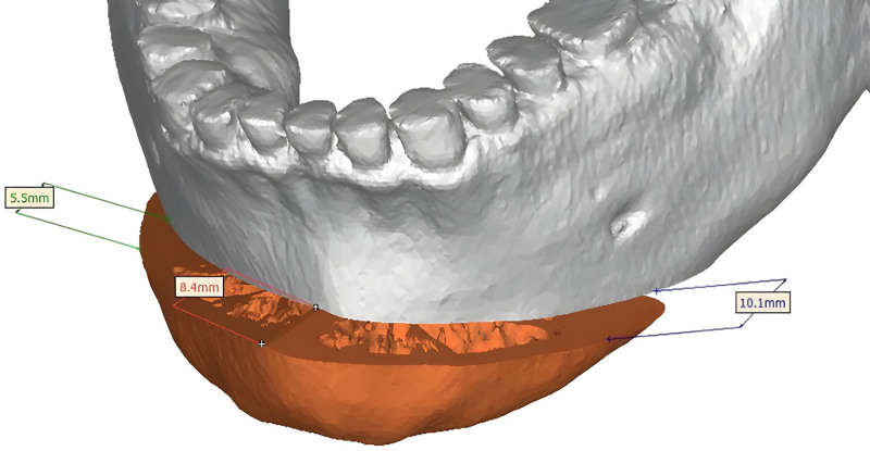
The planes for osteotomy were determined. The studies began with the identification of the mandibular structures, and then the osteotomy line was drawn according to the surgeon’s indications, considering the position of the mental foramen and teeth (Fig. 1 ). Once the virtual planning was validated, the final models exported from the PROPLAN CMF were imported to SolidWorks (Dassault Systèmes, Vélizy-Villacoublay, France) to model the cutting and repositioning guides using a parametric feature-based approach.
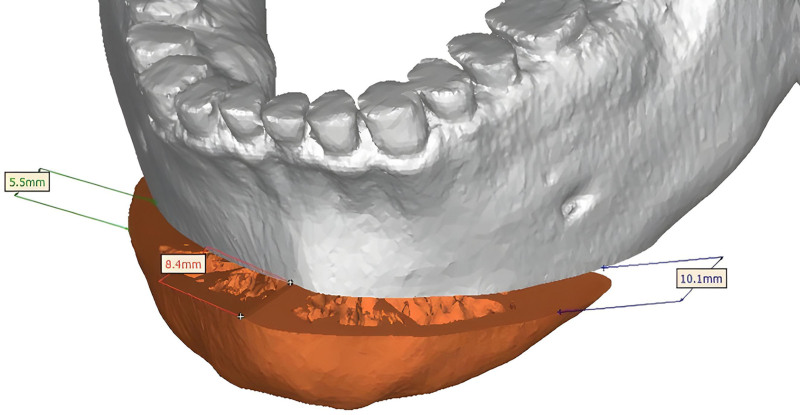
Fig. 1. Virtual osteotomy.- Guide design: After defining the thickness of the mandibular plane, the solid mandible was subtracted to create a thick osteotomy plane that could be fitted to the outer surface of the mandible. Two types of cutting guides were planned: composite and monobloc. The composite was made up of 2 pieces and planned based on the anatomical fit of the mandible using three anatomical landmarks: the lower interincisive midline and the right and left mandibular borders. The mandible was fixed with 2 holes and 2 screws (Fig. 2 ). The monobloc was made as a single piece and was planned based on the anatomical fit of the mandible using 2 anatomical landmarks: the lower interincisive midline and the lower mandible border. The mandible was fixed with 1 or 2 holes and screws (Fig. 3 ).
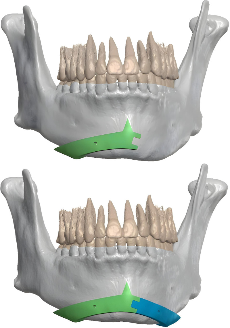
Fig. 2. Virtual composite cutting guide.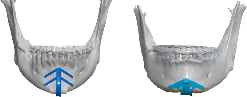
Fig. 3. Virtual monobloc cutting guide.The repositioning guides were planned for 1 piece. They were fixed with 1 or 2 screws using the same holes as the cutting guides (Figs. 4 ).
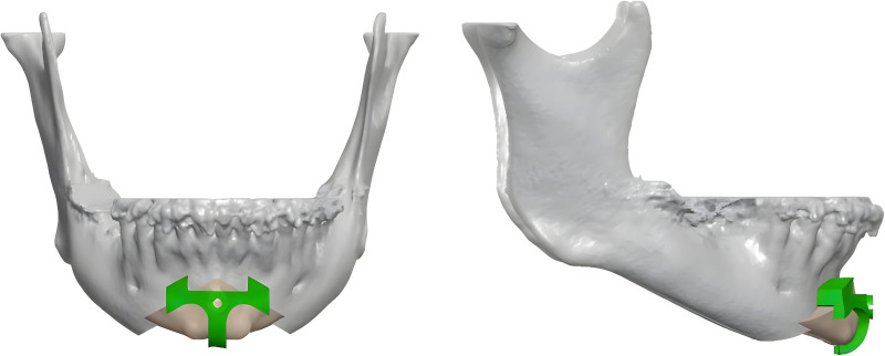
Fig. 4. Virtual repositioning guide.- Manufacturing the surgical cutting guides: The virtual cutting guides for genioplasty and repositioning guides were exported as STL files and applied using a 3D printer. In our in-hospital 3D laboratory, printing was performed using a 3D printer (Formlabs, Somerville, MA). BioMed Clear Resin (Formlabs), a transparent and rigid 3D-printing material with biocompatibility, was selected for printing. Parts printed with BioMed Clear Resin are compatible with common sterilization methods.
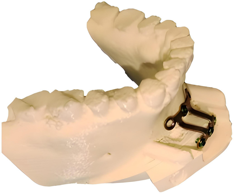
Fig. 5. Printed model with prebending plate.
The procedure was performed under general anesthesia. Ten minutes before surgery, local anesthesia was given through bilateral nerve block injections and infiltrative anesthesia of an anesthetic solution (articaine) that contained a vasoconstrictor (epinephrine), in a ratio of 40 mg/mL of articaine with 0.005 mg/mL of epinephrine, was administered to the alveolar region between both lower premolars and to the mentocervical region. Subsequently, an incision was made in the alveolar mucosa at the first premolar at the level of the vestibular mucosa, at least a couple of millimeters below the adherent gingiva, for proper placement of sutures at the end of surgery. The periosteum was then elevated off the chin bone and the exit of the mental nerves was identified and dissected.
When performing simple osteotomies, a minimally invasive approach can be carried out by means of a longitudinal incision, which allows the osteotomy to be performed safely thanks to the guide, thus enabling a faster recovery and preventing myofascial disturbances in this muscle. 21 – 26 Cutting guides, both monobloc and composite ones, allow a minimal incision; controlled detachment of the soft tissues; creation of a “surgical tunnel”; and, finally, controlled osteotomy of the chin through the use of piezosurgery (Mectron Medical, Carasco, Italy).
After reaching the surgical plane, a customized cutting guide was positioned and fixed for virtual planning. Osteotomy was entirely guided by the splint and performed using piezosurgery.
After the osteotomy, the fragment was fixed to the customized plate using predrilled screw holes in the splint, which facilitated and ensured perfect positioning and fixation of the chin in all 3 dimensions.
After thorough hemostasis, the incision was closed in 2 planes to ensure proper muscle adaptation. Irrigation with physiological saline or chlorhexidine was performed, and a compression dressing was placed on the chin for 3 days to reduce edema and potential postoperative hematoma. Postoperative medications were limited to antibiotics, anti-inflammatory drugs, and corticosteroids for 5 days.
Morphometric analysis was performed by comparing 3D models of the planned and postoperative mandibles.
To perform the analysis, a set of landmarks and related distances was defined as follows:
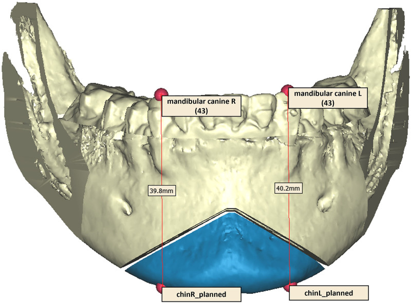
Fig. 6. Virtual distance between the cusp of 43 and 33 and the lower edge of the chin.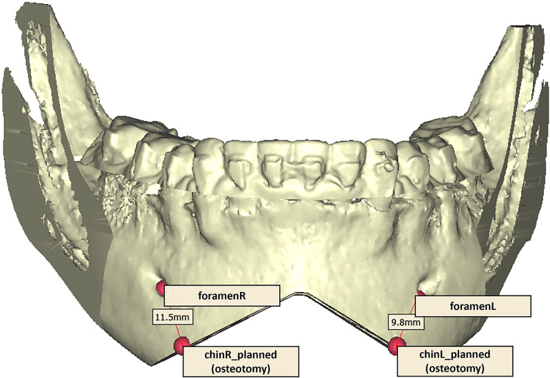
Fig. 7. Virtual distance between the mental foramen landmarks and the osteotomy.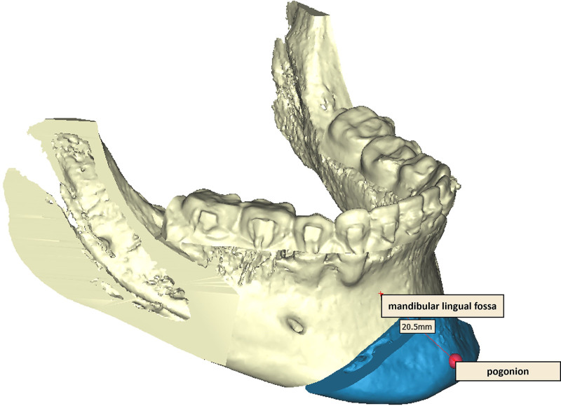
Fig. 8. Virtual distance between mandibular lingual fossa and pogonion.The same set of distances, summarized as 43-chinR, 33-chinL, foramen-chinR, foramen-chinL, and lingual fossa-pog, was computed for the 12 case studies in both planned and postoperative 3D models. The difference in absolute values was then calculated for the accuracy analysis (Table 2 ).
Table 2. Differences Between the Selected Distances to Evaluate Accuracy, Reported for Each Patient, Mean, and SD (σ)
| Patient No. | Δ 43-chinR (mm) | Δ 33-chinL (mm) | Δ lingual fossa-pog (mm) | Δ foramenR-chinR (mm) | Δ foramenL-chinL (mm) |
|---|---|---|---|---|---|
| 1 | 2.8 | 1.8 | 3.3 | 1 | 1 |
| 2 | 1.1 | 0.7 | 0.3 | 1.4 | 1.3 |
| 3 | 1.2 | 1 | 0.6 | 0.4 | 2.5 |
| 4 | 3.1 | 2.4 | 1.8 | 4.2 | 1 |
| 5 | 1.1 | 0.1 | 0.2 | 1.1 | 0.6 |
| 6 | 1.9 | 0.6 | 3.1 | 1.8 | 2.7 |
| 7 | 0.8 | 2 | 3.1 | 2.5 | 3.3 |
| 8 | 0.8 | 1.4 | 1.8 | 1.6 | 0.7 |
| 9 | 0.8 | 1.5 | 3.6 | 0.2 | 0.1 |
| 10 | 0.9 | 1.1 | 1.4 | 0.3 | 0.2 |
| 11 | 1.4 | 1.5 | 2.6 | 1.1 | 0.9 |
| 12 | 0.8 | 0.7 | 1.1 | 1 | 1.3 |
| Mean | 1.39 | 1.23 | 1.91 | 1.38 | 1.30 |
| σ | 0.80 | 0.66 | 1.22 | 1.10 | 1.01 |
Overall, 12 patients were evaluated: 11 were operated on for dysgnathia and 1 underwent surgery for aesthetic reasons. Nine patients underwent combined maxillomandibular surgery (Le Fort I and bilateral sagittal split osteotomy) associated with genioplasty, 1 patient each underwent a Le Fort I operation associated with genioplasty, isolated genioplasty, and secondary genioplasty, respectively. Among the 12 patients, the average duration of genioplasty conducted with piezosurgery was 50.41 minutes. Sliding osteotomy and anteroposterior augmentation were performed in 9 patients. In 2 patients, sliding osteotomy and anteroposterior augmentation were associated with vertical reduction. In 1 patient, asymmetric osteotomy was performed in combination with anteroposterior augmentation. The extent of the anteroposterior augmentation ranged from 3 to 10 mm, with an average of 6.02 mm. The extent of vertical reduction was 3 mm in 1 case and 5 mm in the other.
In 6 cases, the cutting guides were made composite, and in 6 cases, they were made monobloc (Figs. 9 , 10 ). The repositioning guides were all made monobloc (Fig. 11 ). The guides broke in 2 cases; one owing to incorrect positioning and the other because of inadequate thickness.
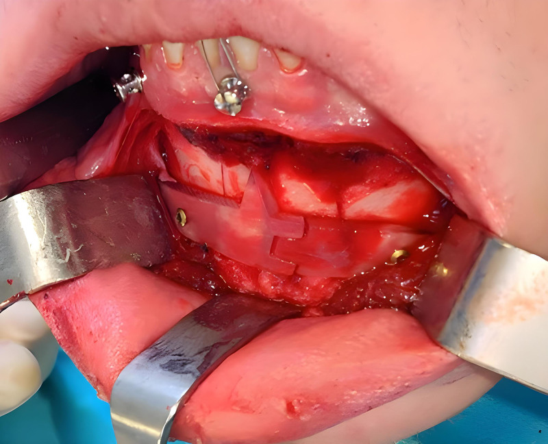
Fig. 9. Intraoperative composite cutting guide.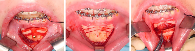
Fig. 10. Intraoperative monobloc cutting guide.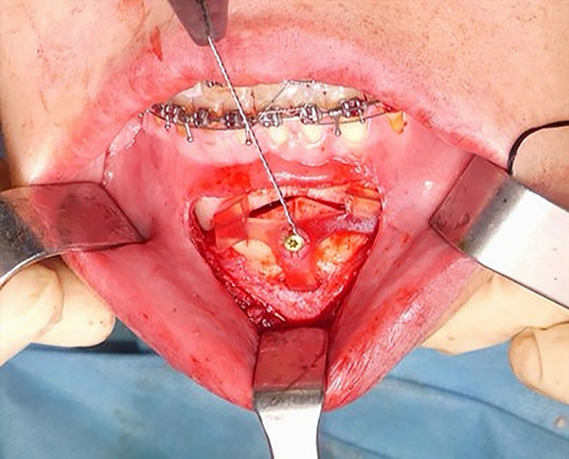
Fig. 11. Intraoperative repositioning guide.No intraoperative adverse events (vascular, nervous, or dental lesions) occurred. Intra- and postoperative bleeding were reduced, as were postoperative edema and hematoma at surgeon’s subjective assessment. Patients were discharged home an average of 2 days after surgery. None of the patients required additional nonsteroidal anti-inflammatory drugs or corticosteroid therapy. Hypoesthesia of the mental nerve occurred in 1 patient during genioplasty. The vitality of dental elements in the interforaminal area was maintained.
From the morphometric analysis comparing 3D models of the planned and postoperative mandibles, we found that the mean and SD were computed to describe the entire set of observations, with a single value representing the center of the data and measuring how spread-out the data were from the mean. Then, to assess and compare the sample data distributions and look for outliers, an individual value plot of multiple variables was created because the sample size was less than 20. As shown in Figure 12 , unlike in a boxplot, each value is displayed separately, which is particularly useful with few observations. An outlier can be identified by analyzing the difference between the foramen-chinR distance on the planned and postoperative 3D models of the mandible, which is probably due to technical difficulties during surgery. As reported in Table 2 , considering the 12 patients analyzed in the current study, the arithmetic averages of the distance between the cusp of the crown of 43 and 33 of the planned and postoperative mandibular edge were 1.39 and 1.23 mm, respectively; the arithmetic average of the distance in the anteroposterior augmentation was 1.91 mm; and the arithmetic averages of the distance between the lower margin of the right and left mental foramen and the upper margin of the planned and postoperative osteotomy were 1.38 and 1.30 mm, respectively. All patients were satisfied with the genioplasty procedure. All operators were satisfied with the cutting and repositioning guides created based on their requests.
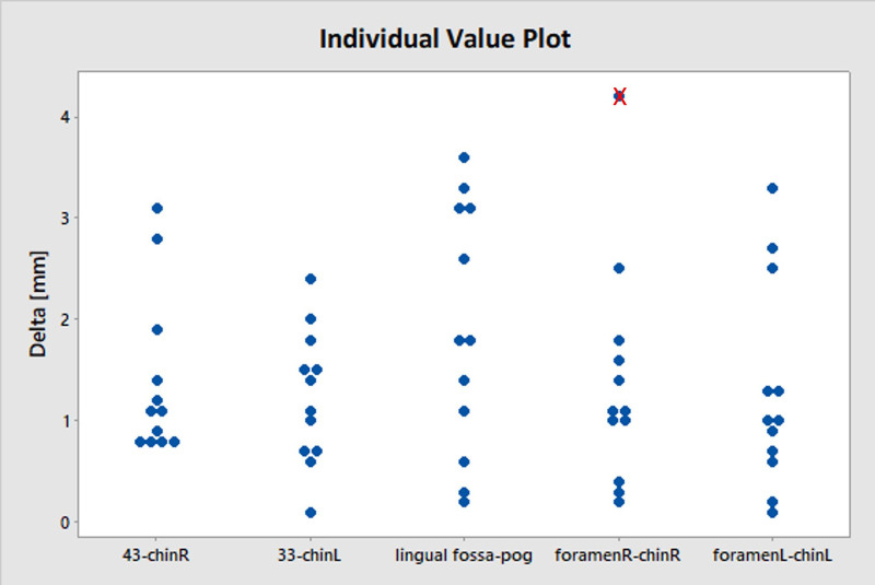
Fig. 12. Individual value plot of multiple variables.
The use of technology has made it possible to create custom devices for maxillofacial surgery. 19 , 20 , 27 , 28 Patients who require mentoplasty expect an improvement in their quality of life by minimizing the intraoperative risks and postoperative complications related to the surgical procedure, resulting in excellent aesthetic and functional results. 29 – 32 Mentoplasty performed using traditional methods is affected by a series of errors owing to limited preoperative planning. Few studies have adequately described the mentoplasty procedure with the aid of 3D technologies in a standardized manner. 15 , 17 , 33 – 37 Mentoplasty performed using rotary instruments causes a series of complications, including irregular osteotomies with sharp bone margins, and the risk of neurovascular and dental lesions. 38 Piezoelectric surgery allows for micrometric and selective cutting of the bone, minimizing the risk of damage to the soft tissues, and creating a cavitation effect that allows for the reduction of intraoperative bleeding with consequent improvement in tissue healing. 39 , 40
We analyzed 12 mentoplasties performed using cutting and repositioning guides designed based on experience and partnership with the 3D Lab at Politecnico of Turin. This procedure is an innovative methodology based on surgeons’ indications. The advantages of mentoplasty conducted with 3D cutting and repositioning guides are greater accuracy and minimal invasiveness compared with the traditional method and the possibility of creating integrated design systems and simulation of the position of the soft tissues during planning. The surgical procedure was simplified, with higher-quality results. The design of these guides has not been described in the literature. 15 , 19 , 35 – 37 , 41 – 45
Cutting guides (composite or monobloc) are designed to facilitate planned osteotomy and have holes for fixation in the same location as the repositioning guides. In vertical reduction, the cutting guides contain the correct size of the symphyseal bone segment, which must be removed to allow impaction of the chin. A monobloc cutting guide allows complex osteotomies with a minimally invasive incision, less periosteum removal, and less postoperative edema. However, a composite cutting guide is simple to place and allows for linear and accurate osteotomy. The repositioning guides are made considering the amount of advancement of the chin and have space to allow the insertion of the osteosynthesis plate that has previously been preblended on the STL model to improve the surgical procedure and reduce operating times. The repositioning guides allow the chin to be fixed in the correct planned position, thereby avoiding asymmetry or unexpected results. 24 The repositioning guides were fixed to the same holes as the cutting guides to determine the controlled movement of the chin bone segment according to the three planes of space. The same procedure could be performed without the aid of the repositioning guides, but with less precision and accuracy. The literature reports that osteotomy of the chin conducted without 3D-printed guides exposes complications in 3%–12% of cases. 29 , 30 , 46 – 48
The mentoplasty procedure described in our study reduces intraoperative bleeding, ptosis of the soft tissues of the chin, and the risk of dental and vascular lesions affecting the mental nerve, owing to the correct osteotomy design and less periosteal removal and traction. 3D-guided mentoplasty conducted with piezosurgery allows for personalized surgical procedures. Consequently, the aesthetic and functional results are more predictable. Three-dimensional-guided mentoplasty allows complex osteotomies such as vertical reduction in cases of asymmetry or in cases of secondary surgical intervention.
Nevertheless, our technique presents some disadvantages: the greater time required for preoperative planning and the need for surgeon training. Certified medical resins occasionally do not resist torsional movements during insertion and fixation. However, the use of resins reduces costs compared with other materials (eg, titanium).
The limitations of our study are its small sample size, the absence of a control group (to compare other tools with objective data), the lack of follow-up, and the need to standardize the shape of the guides.
Compared with reciprocating saws, piezosurgery is used in various fields of maxillofacial surgery. The results have been mixed in terms of patient morbidity. Some authors have affirmed that there is no statistically significant difference in the sensitivity of the labiomental area regarding the instrument used to perform the osteotomy. 49 In contrast, functional sensory recovery of the inferior alveolar nerve following sagittal split osteotomy may be enhanced when a piezoelectric saw is used to perform cortical cuts. 50 According to others, piezosurgery is an effective method for minimizing intraoperative blood loss; operative duration; and postoperative pain, edema, and patient morbidity, and increasing patient satisfaction. 51 , 52 Conventional burrs and microsaws prolong the duration of facial edema. 53 Other authors have affirmed that the choice of the osteotomy tool does not influence the rate of bad fractures. 54 The results of our study encourage the use of 3D cutting and repositioning guides with piezosurgery, not only in the execution of mentoplasties, but also in the daily practice of maxillofacial surgery.
Mentoplasty performed with 3D-printed guides and piezosurgery is simple, accurate, and safe; has excellent functional and aesthetic results; and avoids complications. The costs involved in the creation of the guides are reduced by using 3D printers by approximately €400–€800 per case compared with those outsourced to third-party companies. Cost reduction was calculated based on an average of those provided by major companies.
This process reduces healthcare system costs, allowing access to mentoplasty procedures by a greater number of surgeons (including neophytes), in a multidisciplinary manner, that is, involving engineers.
The authors have no financial interest to declare in relation to the content of this article.
声明:中文精读 · 仅供学术参考。内容来自公开文献检索,不代表本人观点,不构成诊疗建议。 医疗美容需在正规医疗机构由执业医师实施。
基于相同主题推荐 · 继续深挖
12例需颏成形者采用术前CT虚拟手术规划(VSP)联合压电手术(piezosurgery)与3D打印生物相容树脂导板,平均颏成形手术时长50.41分钟。
前瞻性研究纳入 25 例 21–65 岁女性,接受 Cleviel Volume 透明质酸(1.0 mL,22 mg/mL)的 11 点注射(11-PIT)并联合高频超声血管定位进

本研究含2项随机双盲安慰剂对照人体临床试验:19名35岁以上亚洲男性(均龄42±10)使用含1%活性复合物乳膏7天,总毛孔面积较基线下降-30.2%(安慰剂-12.6%)。