前瞻性个体内split-face组织学比较:聚-DL-乳酸(PDLLA)与羟基磷灰石钙(CaHA)皮肤焕活剂——基于干细胞标志物的研究(Plast Reconstr Surg Glob Open)
2026年10月22日29 min read
Plastic and Reconstructive Surgery Global Open
Plastic and Reconstructive Surgery Global Open
结论与实践要点
聚-DL-乳酸(PDLLA)与羟基磷灰石钙(CaHA)均为日益广泛用于皮肤焕活的注射类生物刺激剂。尽管二者均促进组织再生,但其人体组织学层面的差异(尤其是血管、胶原与弹性纤维反应)尚缺乏系统比较。本研究旨在通过个体内split-face设计,在真实人体组织中对比两种制剂的再生特征,为个体化治疗策略提供参考。
本研究为机构审查委员会批准的前瞻性split-face试点研究,纳入3例女性受试者(共6侧面部),一侧注射PDLLA、对侧注射CaHA,于注射后3个月在拉皮术中取对应耳前/颞部活检,采用H&E、Herovici、Verhoeff–Van Gieson染色及CD31/CD34免疫组化,并辅以33 MHz超声与表面粗糙度分析及GAIS评分。结果显示:毛细血管计数PDLLA侧(93,均15.5/片)高于CaHA侧(81,均13.5/片),CD31阳性像素PDLLA(23.6)高于CaHA(16.4);而弹性纤维密度CaHA(165.8±5.54条/视野)高于PDLLA(146.2±5.40),约高13%,CD34表达CaHA(51.4像素)亦高于PDLLA(45.4像素)。超声示两组真皮厚度均显著增加(CaHA 1.027→1.048 mm,P=0.012;PDLLA 1.029→1.056 mm,P=0.0077);粗糙度Ra均显著下降(CaHA 5.10→3.62、PDLLA 5.43→3.00 ×10⁻³mm)。GAIS评分PDLLA侧略高但无统计学差异。
临床意义在于:PDLLA倾向促进血管生成与胶原重塑,CaHA倾向促进弹性生成、祖细胞活化与早期组织加固,二者可视为互补而非互斥,支持按求美者需求个体化选择。但本研究样本量仅3例、随访仅3个月,且缺乏功能性评估(如应力下弹性、灌注),统计把握度有限,结论应审慎解读,仍需更大规模、更长周期研究以明确其临床定位。
Background:Poly-d,l-lactic acid (PDLLA) and calcium hydroxylapatite (CaHA) are injectable biostimulators increasingly used as skin boosters. Although both promote tissue regeneration, their comparative histological effects in humans remain poorly characterized, particularly regarding differential vascular, collagenous, and elastinrelated responses that may influence individualized treatment strategies and clinical decision-making.
Methods:This institutional review board–approved prospective split-face pilot study included 3 female patients (6 cheeks) who received PDLLA injection on one side and CaHA injection on the contralateral side. Biopsies were obtained 3 months after injection from corresponding preauricular or temporal facial regions during the facelift procedure for histological and immunohistochemical evaluation, including hematoxylin and eosin, Herovici, Verhoeff–Van Gieson, and CD31/CD34 immunostaining. Ultrasound (33 MHz) and surface roughness analysis were also performed, alongside Global Aesthetic Improvement Scale scoring.
Results:Both PDLLA and CaHA demonstrated regenerative activity, though their predominant effects differed. PDLLA was associated with greater angiogenesis (capillary counts and CD31 staining), fibroblast activation, and early collagen remodeling. CaHA exhibited higher elastin fiber density (13% increase by Verhoeff–Van Gieson staining), stronger CD34 expression, and more pronounced early dermal thickening on ultrasound. Both agents improved clinical skin roughness and Global Aesthetic Improvement Scale scores without major adverse events.
Conclusions:PDLLA and CaHA skin boosters each demonstrated distinct yet beneficial biostimulatory profiles in human tissue. PDLLA favored angiogenesis and collagen remodeling, whereas CaHA promoted elastogenesis, progenitor cell activation, and early tissue reinforcement. These findings highlight the complementary roles of PDLLA and CaHA and underscore the need for larger, long-term studies to refine their clinical applications.
Takeaways
Question: What are the comparative histological regenerative effects of poly-d,l-lactic acid (PDLLA) and calcium hydroxylapatite (CaHA) skin boosters in human facial tissue?
Findings: In this prospective, intraindividual, split-face pilot study of 3 patients undergoing face lift surgery, both treatments demonstrated regenerative activity. PDLLA showed greater fibroblast activation, CD31 expression, and microvascular responses, whereas CaHA demonstrated greater elastin fiber density and CD34 expression, suggesting distinct histological regenerative profiles.
Meaning: Both PDLLA and CaHA demonstrated regenerative potential in facial tissue, with each showing distinct histological strengths that may support their use as biostimulatory skin boosters.
Facial rejuvenation and scar revision are fundamental goals in aesthetic and reconstructive dermatology. 1 Among the current nonsurgical modalities, injectable biostimulators such as poly-d,l-lactic acid (PDLLA) and calcium hydroxylapatite (CaHA) have emerged as effective agents for inducing neocollagenesis, enhancing dermal quality, and improving textural irregularities. 2 , 3 In particular, the extent to which these injectables influence angiogenesis, elastogenesis, fibroblast activity, and stem cell marker expression remains underinvestigated. 4 – 6 CD31 (PECAM-1) is involved in endothelial cell adhesion and vascular remodeling, serving as a surrogate for angiogenic activity. 7 CD34 marks both vascular progenitor cells and preadipocytes, contributing to both neovascularization and adipogenesis. 8 Additionally, elastin fiber remodeling and fibroblast activation are crucial indicators of dermal regeneration. 9
This study was designed to compare the histological and structural outcomes of CaHA and PDLLA injection using discarded facelift tissue. We hypothesized that both agents would enhance vascularization, fibroblast activation, elastin synthesis, and dermal thickness, but with potentially distinct histological signatures.
This controlled, randomized clinical study included 3 adult patients undergoing elective facelift surgery. To directly compare the histological effects of injectable PDLLA (Juvelook Volume, VAIM Co., Seoul, Korea) and CaHA (DCLASSY,CG BIO Co., Seoul, Korea), each subject received PDLLA on the left side of the face and CaHA on the right side. Biopsies were obtained 3 months after injection from corresponding preauricular or temporal facial regions during the facelift procedure. The harvested tissue, which would otherwise be discarded, was used for histological evaluation with informed consent. The study protocol received institutional review board approval (P01-202404-02-009), and all procedures were conducted in accordance with ethical standards, ensuring participant anonymity and voluntary participation.
PDLLA was prepared by mixing 1 vial with 16 mL of saline, from which 8 mL was injected into the left facial side. CaHA (0.8 mL) was diluted with 8 mL of saline, and the full volume was injected into the right side of the face. Injections were delivered using a 25G, 5-cm blunt cannula at the dermis–subcutaneous interface via 2 entry points per side (malar and oral commissure areas). The distribution aimed to achieve uniform subcutaneous delivery across the face for each material before biopsy and histological analysis.
Histological quantification was performed using a combined approach—manual pathology review, ImageJ-based semiautomated analysis, and artificial intelligence (AI)–assisted evaluation—to improve accuracy, reproducibility, and objectivity beyond conventional methods.
To assess cellular activation in the dermal layer, hematoxylin and eosin (H&E)–stained tissue sections were evaluated under both high-power (×400) and low-power (×20) magnification. AI (ChatGPT-4.0) 10 was trained as activated fibroblasts were defined as spindle-shaped cells with abundant cytoplasm and oval-shaped nuclei, whereas resting fibrocytes were identified as smaller cells with condensed nuclei and minimal visible cytoplasm. 11 – 14 Total cell numbers were calculated for both PDLLA- and CaHA-treated tissues. The sum of activated fibroblasts and resting fibrocytes was recorded and compared between groups to evaluate the differential effects of each material on fibroblast activation status.
To assess neovascularization following injection of CaHA and PDLLA into the subcutaneous fat layer, histologic analysis was performed using H&E-stained sections at 20× magnification. Two representative slides per subject were selected, and capillaries were counted as vascular lumens containing 1–3 red blood cells using the AI. 15 – 17
To evaluate elastogenesis, elastin fibers were visualized using Verhoeff–Van Giepson (VVG) staining, which stains elastic fibers black. Elastin fibers were quantified at ×200 magnification using an automated image analysis using AI. The number of discrete black-stained elastin fibers was counted in 6 slides each from, CaHA-injected and PDLLA-injected tissue to allow direct comparison of elastin density between the 2 treatment modalities. 18
To objectively assess the expression of CD31 7 and CD34 8 stem cell markers, immunohistochemically stained tissue sections were analyzed using AI-assisted digital image analysis. High-resolution whole-slide images were captured at ×200 magnification, and each image field consisted of exactly 1,774,216 pixels. The AI algorithm was trained to distinguish positively stained brown pixels indicating CD31 or CD34 expression from background tissue and nonspecific blue staining. All brown pixels within each image were automatically counted, and their total was recorded.
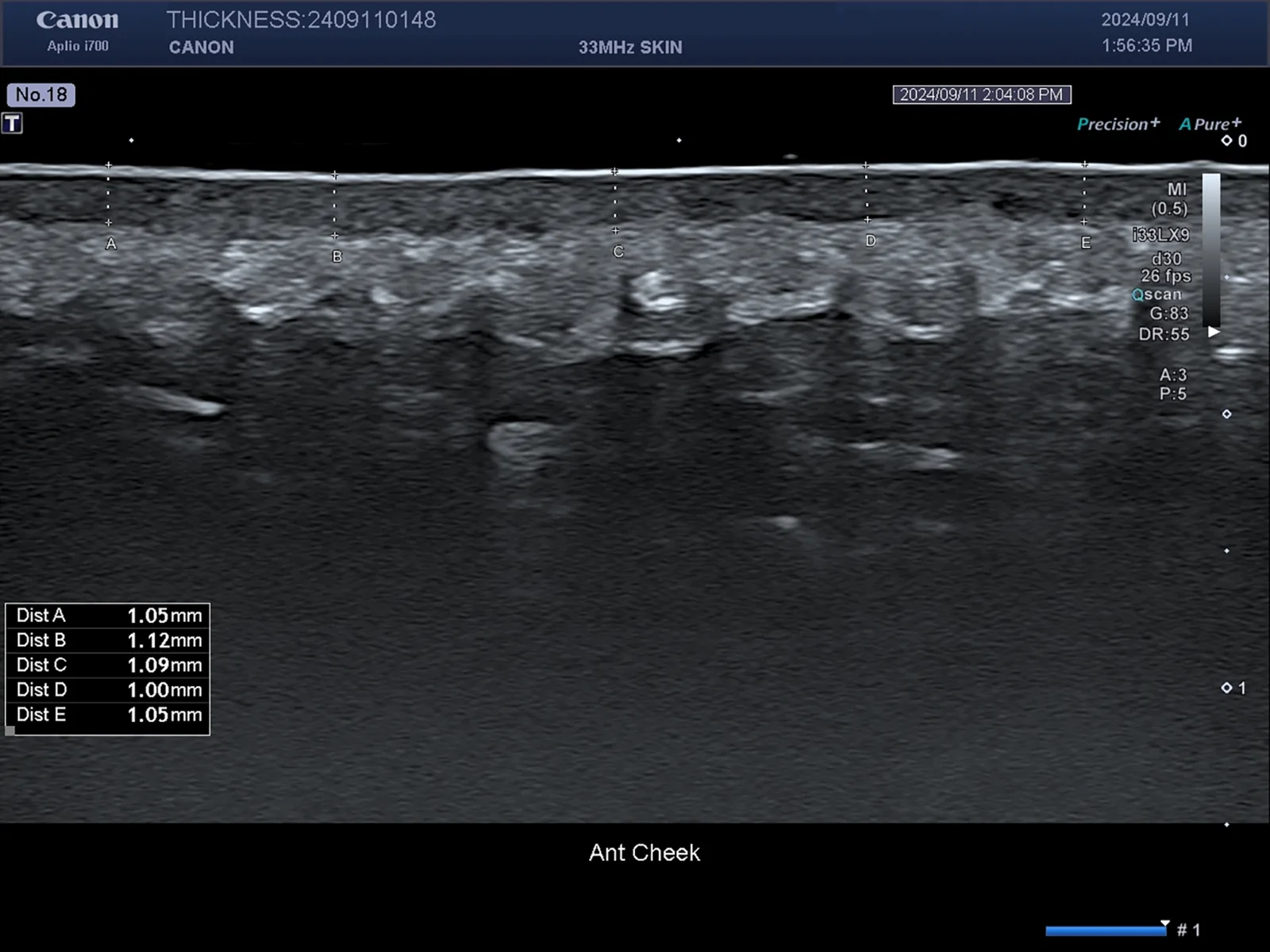
Skin thickness was measured using an Aplio i700 (Canon, Co., Japan) ultrasound device with a 33-MHz linear probe. Measurements were taken 10 sites from the left and right anterior cheek. Assessments were conducted at 2 time points, before injection and at 3 months, to evaluate changes in dermal and subcutaneous tissue thickness over time (Fig. 1 ).
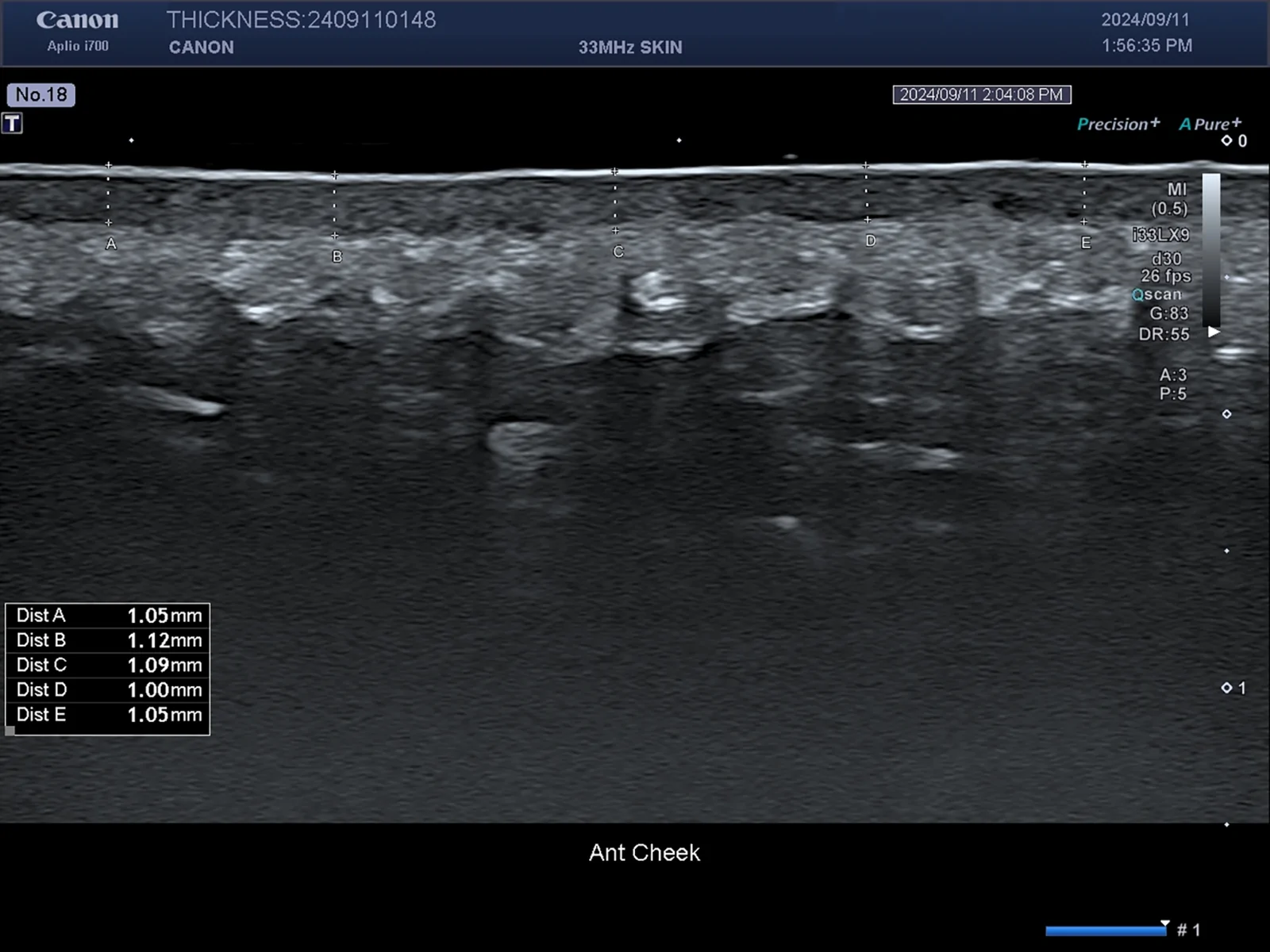
Fig. 1. Ultrasonographic measurement of skin thickness before injection. Representative 33-MHz ultrasound image of the right anterior cheek before injection, demonstrating baseline dermal and subcutaneous tissue thickness, yielding an average skin thickness of 1.06 mm. These baseline values were compared with follow-up scans at 3 months postinjection to evaluate the dermal remodeling effects of PDLLA and CaHA.注射前皮肤厚度超声测量示例(33 MHz),显示基线真皮与皮下组织厚度,平均皮肤厚度1.06 mm,作为3个月随访对照基线。
Skin roughness was quantitatively assessed in 3 patients at 5 left and 5 right sites each, using Gwyddion software (v2.62). 19 , 20 Line scan profilometry was performed along the submalar line from the tragus to the oral commissure, a region susceptible to fine wrinkles. Average roughness (Ra) was calculated to evaluate fine wrinkle density and surface irregularities, enabling comparative analysis of dermal remodeling between PDLLA- and CaHA-treated areas (Fig. 2 ).
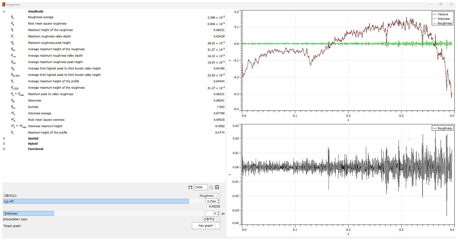
Fig. 2. Surface roughness analysis using Gwyddion software (version 2.62). Profilometric evaluation of the anterior cheek region using 2D line scan data extracted with Gwyddion software. The graphs display surface texture (blue), waviness (red), and high-frequency roughness components (green) over a 0.4-mm horizontal distance. Roughness parameters including average roughness (Ra = 3.398 µm) and root-mean-square roughness (Rq = 4.906 µm) were computed to quantify micrometric variations in skin surface contour.使用Gwyddion软件的表面粗糙度分析,显示前颊区轮廓扫描的纹理、波纹与高频粗糙度分量,基线Ra=3.398 µm、Rq=4.906 µm。
Patient satisfaction was evaluated at 3 months posttreatment using the Global Aesthetic Improvement Scale (GAIS), a validated 5-point subjective scale assessing the degree of improvement compared to baseline. Each of the 3 patients independently rated improvements in 4 aesthetic domains: skin texture, fine wrinkle reduction, skin tone, and skin elasticity, separately for the CaHA-treated and PDLLA-treated sides of the face.
This study included only 3 patients due to the invasive nature of facial biopsy. Although the sample size was limited, combining patient-reported outcomes with objective histologic and instrumental data still provides meaningful preliminary evidence.
Quantitative histological analysis was conducted to compare the activation status of fibroblasts between CaHA- and PDLLA-treated tissues using H&E-stained sections. At high magnification (×400), PDLLA-treated samples exhibited a significantly greater number of activated fibroblasts (30.88 ± 2.04) compared to CaHA-treated samples (14.00 ± 2.36). Conversely, CaHA-treated tissues showed a higher number of resting fibrocytes (13.00 ± 1.41) than PDLLA-treated samples (12.50 ± 1.87), although the difference was modest.
At low magnification (×20), automated cell counts across 6 representative fields further confirmed this trend. PDLLA-treated tissues demonstrated an average of 595.33 ± 195.21 activated fibroblasts per field, markedly exceeding the 475.83 ± 134.35 observed in CaHA-treated tissues. Interestingly, the number of resting fibrocytes was also higher in PDLLA-treated samples (612.00 ± 68.40) compared to CaHA (408.50 ± 48.20), possibly reflecting an overall increase in total fibroblast lineage cells within the PDLLA group.These findings suggest that PDLLA may exert a stronger stimulatory effect on fibroblast activation and recruitment than CaHA, potentially contributing to enhanced extracellular matrix remodeling and dermal regeneration (Table 1 ; Figs. 3 , 4 ).
Table 1. Quantitative Comparison of Activated Fibroblasts and Resting Fibrocytes in PDLLA- and CaHA-treated Tissue
| Magnification | Fibroblast-activated CaHA | Fibrocyte Resting CaHA | Fibroblast-activated PDLLA | Fibrocyte Resting PDLLA |
|---|---|---|---|---|
| X400 | 16 | 13 | 28 | 11 |
| 11 | 14 | 31 | 13 | |
| 12 | 15 | 31 | 15 | |
| 15 | 12 | 33 | 10 | |
| 13 | 13 | 33 | 14 | |
| 17 | 11 | 29 | 12 | |
| Total | 84 | 78 | 185 | 75 |
| Average (SD) | 14.00 ± 2.36 | 13.00 ± 1.41 | 30.88 ± 2.04 | 12.50 ± 1.87 |
| X20 | 554 | 421 | 397 | 635 |
| 573 | 419 | 855 | 688 | |
| 499 | 320 | 346 | 673 | |
| 514 | 401 | 647 | 542 | |
| 208 | 465 | 594 | 614 | |
| 507 | 425 | 733 | 520 | |
| Total | 2855 | 2451 | 3575 | 3672 |
| Average (SD) | 475.83 ± 134.35 | 408.50 ± 48.20 | 595.33 ± 195.21 | 612.00 ± 68.40 |
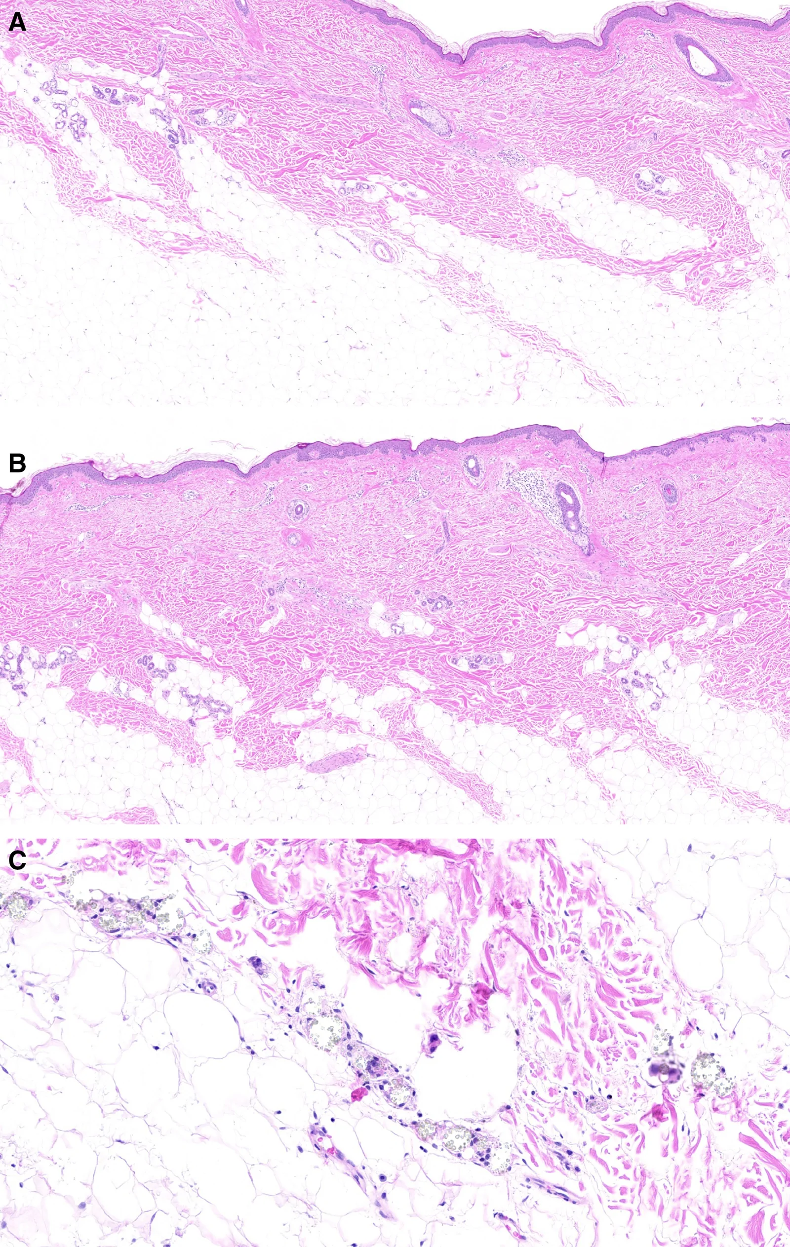
Fig. 3. Histologic comparison (H&E) of dermal and subcutaneous tissue remodeling between PDLLA and CaHA injections. A, In H&E stain from a CaHA-injected facial region (×20), the dermal layer appears moderately thickened with fibroblast activation, and extracellular matrix remodeling is observed. B, PDLLA-injected site (×20) showed pronounced dermal thickening with denser collagen fiber alignment and increased fibroblast-like cellularity. The subcutaneous layer demonstrates enhanced matrix organization with apparent interfascicular fibrous septa, suggesting tissue regeneration. C, In the CaHA group (×200), there were multinucleated foreign giant cells, CaHA microparticles (gray), indicating newly deposited collagen bundles and active fibroblast infiltration, suggestive of regenerative stimulation.H&E染色下PDLLA与CaHA注射后真皮及皮下组织重塑对比:CaHA侧真皮中度增厚伴成纤维细胞活化,PDLLA侧真皮增厚更明显、胶原排列更致密,CaHA侧可见多核巨细胞与微颗粒。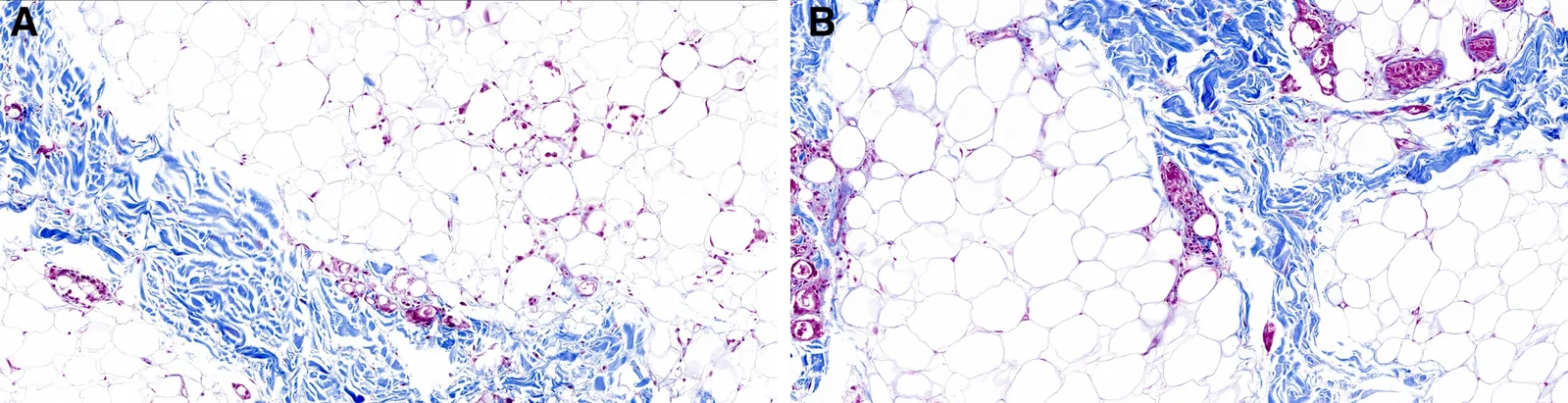
Fig. 4. Comparative histologic evaluation of collagen deposition and adipocyte following injection of CaAH and PDLLA biostimulators with Masson trichrome staining. A, Tissue from a subject injected with CaHA-(DCLASSY) showed dense blue-stained collagen bundles, indicating marked neocollagenesis along the dermal-subcutaneous junction with new healthy adipocytes. B, Tissue from a subject injected with PDLLA (Juvelook Volume) demonstrates moderate collagen deposition with newly made thin blue collagen fibers and healthy adipocytes.Masson三色染色比较CaHA与PDLLA注射后胶原沉积与脂肪细胞:CaHA侧见致密蓝色胶原束与新生成熟脂肪细胞,PDLLA侧为中等胶原沉积与新生的细胶原纤维。
At 3 months postinjection, the CaHA-treated group exhibited a total of 81 capillaries across 6 slides (mean per slide: 13.5, SD: 1.87), whereas the PDLLA-treated group showed a total of 93 capillaries (mean per slide: 15.5, SD: 2.43). Although statistical significance could not be established due to the small sample size, the PDLLA group demonstrated a trend toward higher capillary counts. These findings suggest that both CaHA and PDLLA may enhance microvascular formation in the subcutaneous layer following injection, with slightly greater capillary presence observed in PDLLA-treated tissue (Table 2 ; Figs. 3 , 4 ).
Table 2. Capillary Count per H&E-stained Slide (×20 Magnification) in CaHA- and PDLLA-treated Subcutaneous Tissue
| Capillary (1,2,3) in ×20 H&E Slide | CaHA | PDLLA |
|---|---|---|
| Subject 1 | 11 | 12 |
| Subject 1 | 16 | 15 |
| Subject 2 | 15 | 19 |
| Subject 2 | 14 | 16 |
| Subject 3 | 12 | 14 |
| Subject 3 | 13 | 17 |
| Total | 81 | 93 |
| Average | 13.5 | 15.5 |
| SD | 1.8 | 2.4 |
Quantitative analysis of Verhoeff-stained slides demonstrated that the CaHA-injected tissues exhibited a higher elastin fiber density compared to the PDLLA-injected tissues. The mean number of elastin fibers per ×200 magnification field was 165.8 ± 5.54 for CaHA and 146.2 ± 5.40 for PDLLA. The total count across all evaluated fields was 1005 fibers for CaHA versus 899 fibers for PDLLA, indicating an approximately 13% increase in elastin content in the CaHA-treated regions. This finding suggests that CaHA promotes greater elastogenesis compared to PDLLA (Table 3 ; Fig. 5 ).
Table 3. Quantification of Elastin Fibers in Verhoeff-stained Tissue Sections at ×200 Magnification
| Verhoeff (×200) Elastic Fibers Number | CaHA | PDLLA |
|---|---|---|
| Subject 1 | 157 | 138 |
| Subject 1 | 167 | 150 |
| Subject 2 | 165 | 145 |
| Subject 2 | 168 | 152 |
| Subject 3 | 172 | 146 |
| Subject 3 | 176 | 168 |
| Total fibers | 1005 | 899 |
| Average fibers | 165.8 | 146.2 |
| SD | 5.54 | 5.40 |
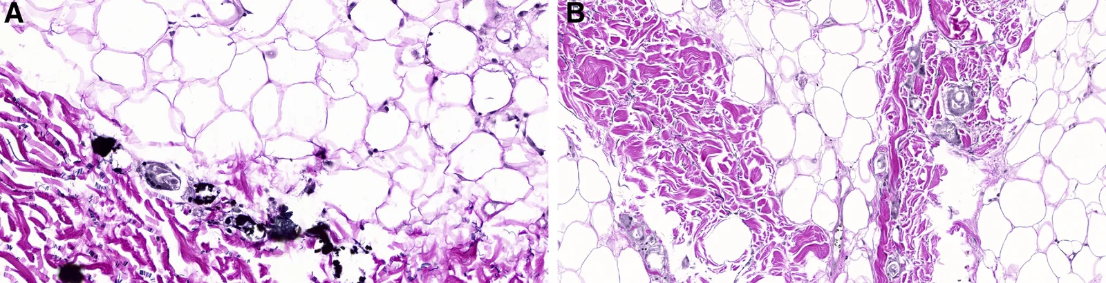
Fig. 5. VVG staining for elastic fiber evaluation at 200× magnification. A, In the DCLASSY-treated tissue highlighted abundance of dense and fragmented elastic fibers (black) within the dermis, showing elastogenesis and reflecting active extracellular matrix remodeling. B, The Juvelook Volume group demonstrated abundance elastic fibers with a more preserved and linear pattern, suggesting elastogenic activity.VVG染色(200×)评估弹性纤维:CaHA侧弹性纤维密集且呈碎片化,PDLLA侧弹性纤维丰富且排列更规则线性,均提示弹性生成活跃。
Quantitative analysis revealed that the CaHA group exhibited an average of 16.4 CD31-positive pixels per ×200 field, corresponding to 4.67 × 10 −3 % of the total image pixels. In contrast, the PDLLA group showed a higher CD31 expression, averaging 23.6 positive pixels, or 1.02 × 10 −2 % of the field. Although both treatments promoted neovascularization to a certain extent, the PDLLA group demonstrated a more pronounced increase in CD31-positive endothelial marker expression, suggesting relatively greater angiogenic activity at 3 months postinjection (Table 4 ; Fig. 6 ).
Table 4. Quantitative Analysis of CD31 Expression at ×200 Magnification Following CaHA and PDLLA Injection
| CD31 Expression (×200) | CaHA | PDLLA |
|---|---|---|
| Subject 1 | 21 | 32 |
| Subject 1 | 15 | 17 |
| Subject 2 | 21 | 37 |
| Subject 2 | 10 | 18 |
| Subject 3 | 15 | 14 |
| Subject 3 | 13 | 48 |
| Total CD31 pixel | 95 | 166 |
| Average pixel | 16.4 | 23.6 |
| SD | 4.66 | 10.21 |
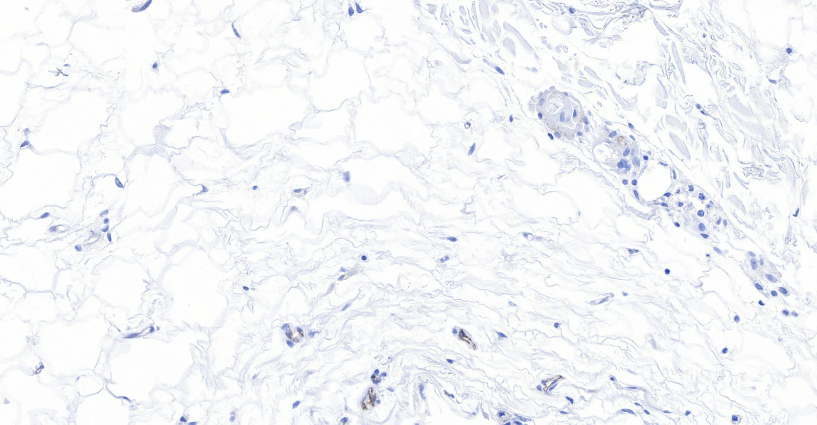
Fig. 6. CD31 immunohistochemical staining of subcutaneous tissue in the CaHA-treated group (×200). CD31-positive endothelial cells (brown staining) were observed within the dermis and subcutaneous tissue, indicating the presence of newly formed microvessels along the vascular lumina and interstitial zones, reflecting angiogenic activation following sublethal stimulation.CaHA组皮下组织CD31免疫组化(200×),棕色阳性内皮细胞见于真皮及皮下,提示新生微血管形成与血管生成激活。
In the CaHA group, CD34-positive staining accounted for an average of 51.4 pixels per ×200 field, corresponding to approximately 0.00290% of the total image area (1,774,216 pixels per field). The PDLLA group exhibited a slightly lower expression, averaging 45.4 pixels per field, or 0.00256% of the total field. Despite the small proportion of CD34-positive area overall, CaHA treatment resulted in a modestly higher level of CD34 expression, which may reflect increased presence or activation of preadipocytes and endothelial progenitor cells within the subcutaneous tissue microenvironment (Table 5 ; Fig. 7 ).
Table 5. Quantitative Analysis of CD34 Expression at ×200 Magnification Following CaHA and PDLLA Injection
| CD34 Expression (×200) | CaHA | PDLLA |
|---|---|---|
| Subject 1 | 59 | 34 |
| Subject 1 | 34 | 41 |
| Subject 2 | 68 | 31 |
| Subject 2 | 45 | 52 |
| Subject 3 | 51 | 69 |
| Subject 3 | 59 | 41 |
| Total CD34 pixel | 316 | 268 |
| Average pixel | 51.4 | 45.4 |
| SD | 13.01 | 15.46 |
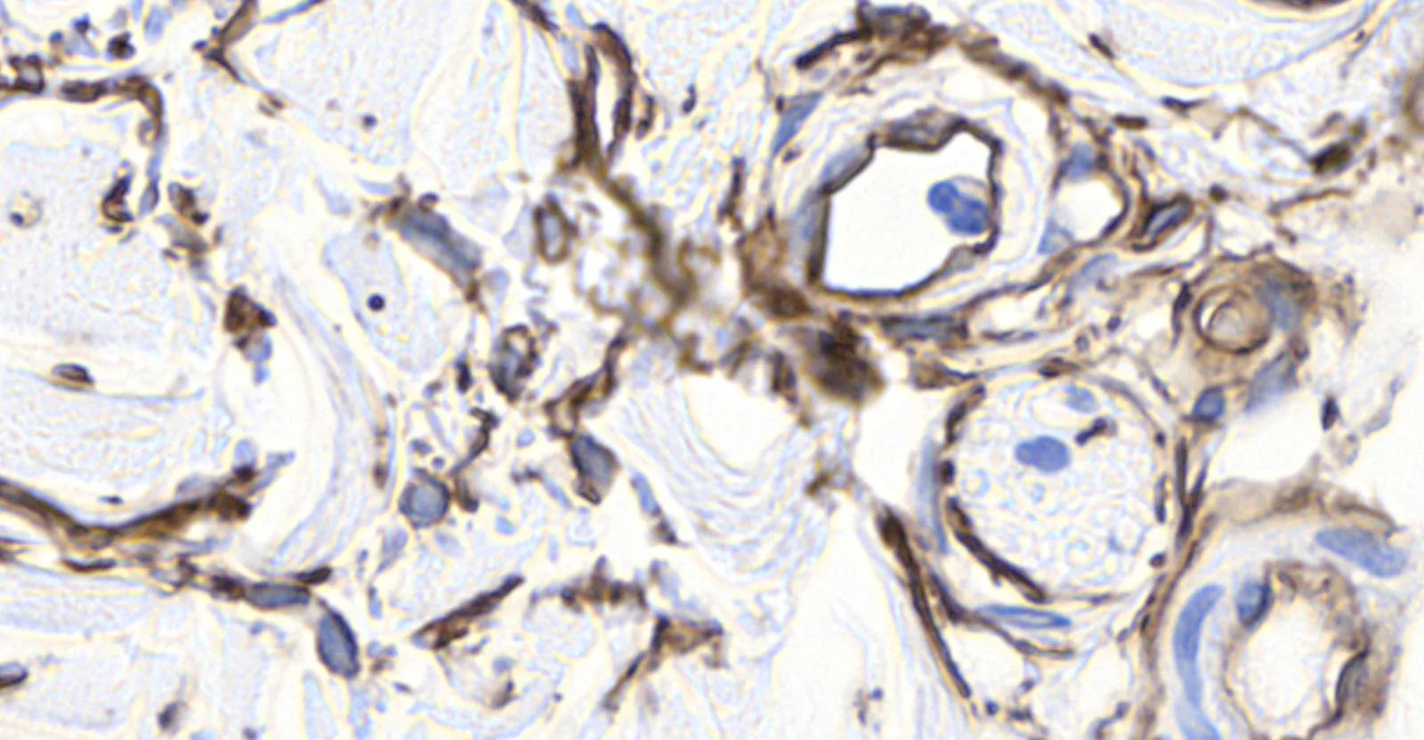
Fig. 7. High-power view (×400) of CD34 immunohistochemical staining in CaHA-treated group. CD34-positive cells (brown staining) were visualized in perivascular and interstitial regions of the subcutaneous layer, indicating activation or recruitment of adipose-derived stem/progenitor cells and endothelial progenitor cells. The CaHA-treated tissue demonstrates increased CD34 expression along the microvascular and stromal interfaces, consistent with enhanced adipogenic and angiogenic responses, suggesting that CaHA injection may stimulate regenerative cellular populations within the subcutaneous microenvironment.CaHA组CD34免疫组化高倍镜(400×),棕色阳性细胞见于血管周围及间质,提示脂肪来源干细胞/祖细胞与内皮祖细胞活化或募集增加。
In the CaHA group, mean dermal thickness increased from 1.027 ± 0.047 mm pretreatment to 1.048 ± 0.048 mm at 3 months posttreatment. A paired t test demonstrated a statistically significant increase in dermal thickness (t = 2.67, P = 0.012) ( P > 0.001).
In the PDLLA group, mean dermal thickness increased from 1.029 ± 0.070 mm pretreatment to 1.056 ± 0.069 mm at 3 months. This increase was also statistically significant. (t = 2.87, P = 0.0077) ( P > 0.001).
When comparing the degree of thickness change between the 2 groups, both injectables demonstrated similar levels of dermal thickening. Both the PDLLA- and CaHA-treated side showed a slight increase with no stastical significance ( P > 0.001), suggesting a regeneration in the dermis and volumizing response in the fat layer. These findings support the hypothesis that both CaHA and PDLLA stimulate dermal remodeling, with PDLLA potentially inducing slightly more sustained fibroblast activation and matrix production over time (Fig. 1 ).
Skin surface roughness was quantitatively evaluated using the Gwyddion software. A total of 30 sites for 3 participants underwent profilometric surface analysis along the submalar line (from tragus to mouth corner) at baseline and 3 months following injection with either CaHA on the right side or PDLLA on the left. The average pretreatment roughness (Ra, ×10 −3 mm) was 5.10 ± 0.92 for the CaHA side and 5.43 ± 0.95 for the PDLLA side. After 3 months, roughness significantly decreased to 3.62 ± 0.58 on the CaHA-treated side and 3.00 ± 0.53 on the PDLLA-treated side (Fig. 2 ).
Paired t tests demonstrated statistically significant improvements in surface smoothness for both treatment modalities (CaHA: P = 3.47 × 10 −8 , PDLLA: P = 1.98 × 10 −13 ), indicating a significant reduction in fine wrinkles and surface irregularities after both treatments. Notably, the PDLLA side exhibited a slightly greater absolute reduction in roughness compared to the CaHA side. Although reductions in skin surface roughness were observed on both the CaHA- and PDLLA-treated sides, the limited sample size precludes definitive statistical significance.
All 3 patients showed visible improvements in skin texture, wrinkles, tone, and elasticity after both CaHA and PDLLA. GAIS scores were slightly higher on the PDLLA-treated side (texture 4.67 versus 4.0, wrinkles 5.0 versus 4.33, tone 4.67 versus 4.33, and elasticity 5.0 versus 4.67). Because the sample size was limited to 3 patients, these differences are not statistically significant.
Fibroblasts, the primary cells responsible for synthesizing extracellular matrix components like glycosaminoglycans and hyaluronic acid, were quantified in this study. An increase in fibroblast count is typically associated with enhanced skin health and regeneration. 21 Beyond their quantity, changes in fibroblast morphology, such as increased cell size and elongated shape, often indicate heightened metabolic activity and collagen production. 22 The presence of specific stem cell markers correlates with fibroblast activity and skin regeneration. For instance, CD34-positive endothelial progenitor cells play a role in angiogenesis and can indirectly influence fibroblast function by enhancing vascularization and tissue perfusion. 23 , 24 This marker is crucial for understanding how fibroblasts contribute to skin health and regeneration mechanisms.
The number of newly formed capillaries was quantified to assess the improvement in microvascular density and blood circulation. 25 Increased capillary density indicates enhanced nutrient and oxygen delivery to the tissues, which are essential for cellular metabolism and wound healing. Therefore, monitoring capillary density provides objective evidence of the biological effects of PDLLA and CaHA injections on tissue microcirculation and vascular support. The key stem cell markers associated with neo-angiogenesis are CD34 and CD31. 26 CD34 is used to identify endothelial progenitor cells and preadipocytes. CD31 (PECAM-1) is known as platelet endothelial cell adhesion molecule-1, and is closely associated with endothelial cells in blood vessels. It is important for vascular formation, inflammation regulation, and cellular interactions. 7 , 27
The presence and condition of elastic fibers was observed. 28 , 29 To assess changes in elastic fibers, stains like VVG or Victoria blue can be used. VVG stain specifically highlights elastic fibers in black or dark brown, contrasting with surrounding tissues. 30 Victoria blue stain, on the other hand, stains elastic fibers blue, allowing for a different visual assessment of their presence and distribution. 31 The presence and condition of elastic fibers is closely related to certain stem cell markers involved in tissue elasticity and repair.
CD31 and CD34 are pivotal markers implicated in the regenerative processes following injectable skin booster treatments. CD31, primarily expressed on vascular endothelial cells, contributes significantly to angiogenesis, ensuring sufficient perfusion and nutrient delivery to regenerating tissues. 26 Moreover, its role in modulating inflammatory responses and mediating intercellular adhesion supports coordinated tissue repair and stabilization of the microvascular environment. CD34, a well-recognized marker of endothelial progenitor cells and preadipocytes, not only facilitates neovascularization but also contributes to adipogenesis, restoring volume and structural integrity in facial soft tissues. 32 Together, these markers reflect critical biological mechanisms—vascular remodeling and stromal cell activation—that underlie the clinical improvements observed with CaHA and PDLLA treatments.
Both PDLLA and CaHA injections induced notable histological changes in facial tissues, supporting their efficacy in aesthetic skin improvement. However, PDLLA demonstrated superior performance in key regenerative processes. Quantitative histological analysis revealed that PDLLA-treated tissue showed a greater density of CD31-positive capillaries, suggesting enhanced angiogenesis, which is critical for sustained tissue oxygenation and nutrient delivery. Fibroblast activation was also more prominent in the PDLLA group, as evidenced by the increased presence of activated fibroblasts with oval nuclei and cytoplasmic elongation, leading to greater collagen synthesis. Although VVG staining confirmed a higher count of elastin fibers following CaHA treatment, reflecting enhanced dermal remodeling. Although PDLLA showed relatively stronger fibroblast activation and angiogenesis, CaHA demonstrated superior elastin fiber formation, higher CD34 expression, and significant early dermal thickening. Given its biocompatibility as a calcium-based material and its immediate volumizing effects, CaHA represents a safe and effective option that complements the long-term regenerative effects of PDLLA. Specifically, CaHA-injected tissues exhibited a 13% higher elastin fiber density compared to PDLLA, suggesting a more robust stimulation of elastogenesis that contributes to skin elasticity and prevention of sagging. CaHA also produced a statistically significant increase in dermal thickness even in the short term ( P = 0.012), supporting its role in early clinical improvement and patient satisfaction. In addition, the immediate volumizing capacity of CaHA allows for rapid correction of contour deficiencies and wrinkle reduction, a feature not shared by PDLLA, which mainly relies on delayed collagen stimulation. Taken together, these findings suggest that although PDLLA excels in long-term biostimulation, CaHA provides a unique combination of immediate safety, biocompatibility, and regenerative efficacy, making it a valuable therapeutic option in clinical practice.
This study has several limitations. First, the sample size was limited to 3 subjects due to the invasive nature of tissue biopsy in aesthetic procedures, which may restrict the generalizability of the findings. Second, the observation period was confined to 3 months, and longer-term histological and clinical outcomes were not assessed. Third, although quantitative analysis of angiogenesis, fibroblast activation, and marker expression was conducted, functional evaluations of tissue performance, such as elasticity under stress or vascular perfusion, were not included. Finally, although AI-assisted image analysis enhanced objectivity, potential variability in staining intensity and tissue sectioning could introduce minor inconsistencies. Future studies with larger cohorts, extended follow-up periods, and functional outcome assessments are warranted to validate and expand upon these findings. Nevertheless, this study provides the first direct quantitative histological evidence from human facial tissue demonstrating distinct biostimulatory signatures of PDLLA versus CaHA.
Although AI-based image analysis offers significant advantages in terms of objectivity and reproducibility, it is not without limitations. The current algorithm’s performance is dependent on the quality of training data, and its recognition accuracy may vary across different histological stains or tissue contexts. Unlike human pathologists, AI systems may lack contextual awareness when encountering unexpected artifacts. Therefore, validation against expert consensus remains essential for ensuring reliability. Although AI-assisted quantification demonstrated high internal consistency in this study, further validation against a consensus panel of expert dermatopathologists remains necessary to establish external validity and ensure generalizability across different histologic platforms. 33 – 39
This prospective intraindividual split-face pilot study provides the first direct histological comparison of PDLLA and CaHA skin booster injections using human facial tissue harvested during facelift surgery. Both injectables demonstrated meaningful regenerative activity at 3 months, including improvements in dermal structure, extracellular matrix remodeling, microvascularity, and patient-reported aesthetic outcomes. PDLLA showed greater fibroblast activation, higher CD31-positive endothelial activity, and a trend toward increased microvascular density, supporting its role in sustained angiogenesis and progressive collagen remodeling. In contrast, CaHA demonstrated superior elastin fiber formation, higher CD34 expression, and significant early dermal thickening, suggesting enhanced elastogenesis, progenitor cell recruitment, and early structural reinforcement. CaHA additionally exhibited favorable short-term safety and biocompatibility consistent with its calcium-based composition. As a ceramic-based biomaterial, CaHA appears to promote a more gradual and physiologic regenerative response with relatively mild tissue reactivity, suggesting a lower potential for excessive inflammatory reactions and related adverse events while maintaining effective tissue regeneration. Although limited by the small sample size and short follow-up period, this pilot study establishes foundational histological evidence that PDLLA and CaHA induce distinct regenerative signatures in human tissue. These findings may help refine patient-specific selection of biostimulatory injectables and support future larger-scale studies with extended follow-up and expanded biomarker analysis.
The author has no financial interest to declare in relation to the content of this article.
The author expresses sincere gratitude to Yu Hyun Seung for his expert insight and support in the development of this study.
声明:中文精读 · 仅供学术参考。内容来自公开文献检索,不代表本人观点,不构成诊疗建议。 医疗美容需在正规医疗机构由执业医师实施。
基于相同主题推荐 · 继续深挖
本研究为IRB批准的前瞻性split-face试点研究,纳入3例女性(共6侧面部),一侧注射PDLLA、对侧注射CaHA,于注射后3个月在拉皮术中取对应部位活检行组织学与免疫组化评
前瞻性研究纳入 25 例 21–65 岁女性,接受 Cleviel Volume 透明质酸(1.0 mL,22 mg/mL)的 11 点注射(11-PIT)并联合高频超声血管定位进
回顾性研究纳入23例接受JAMBER(JB)盘绕PDO线鼻部塑形患者(8男15女,平均35.3岁,21–72岁),随访至术后12个月。