1.5 mm 与 3.0 mm 聚焦超声(HIFU)用于韩国成人眉部提升的疗效与安全性:一项前瞻性随机 split-face 研究(《Skin Research and Technology》)
2026年10月22日35 min read
Skin Research and Technology
Skin Research and Technology

结论与实践要点
背景与目的:高强度聚焦超声(HIFU)已被广泛用于非侵入性眉部提升,但同一受试者内不同焦点深度 cartridge 的疗效比较较为缺乏。本研究旨在评估 1.5 mm 与 3.0 mm 两种聚焦超声 cartridges 在韩国成人眉部提升中的疗效与安全性,并开展探索性的深度间比较。
方法学与关键结果:本研究为前瞻性随机 split-face 研究,共 20 名韩国成人接受单次治疗,一侧额部使用 1.5 mm cartridge、对侧使用 3.0 mm cartridge(每侧 225 J),在基线、术后即刻及第 1、4、12 周以标准化照片测量平均眉高(AEH,主要终点)与最大眉高(MEH,次要终点)。结果显示 12 周时 AEH 较基线分别提升 1.76±1.19 mm(1.5 mm)与 1.23±1.33 mm(3.0 mm);经多重校正后两种 cartridges 均满足 AEH 主要疗效标准,而 MEH 仅 1.5 mm 达标(0.57 mm),3.0 mm 未达标(-0.08 mm)。探索性 12 周 AEH 深度间差异无统计学显著性(p=0.116)。12 周随访期内未发生治疗相关不良事件,受试者满意度较高。
临床意义与局限:两种 cartridges 在 12 周内均带来有统计学意义的眉高提升且短期耐受性良好,但研究缺乏假体对照组,仅能说明自身改善而非因果疗效;深度间比较为事后探索性分析且样本量小、置信区间较宽,结果尚无定论,需更大规模、设假对照的研究进一步验证。
BackgroundHigh‐intensity focused ultrasound (HIFU) is widely used for noninvasive eyebrow lifting, but within‐subject comparisons of cartridge focal depths are scarce.
ObjectiveTo evaluate the efficacy and safety of 1.5‐ and 3.0‐mm cartridges for eyebrow lifting in Korean adults, with an exploratory between‐depth comparison.
MethodsIn this randomized split‐face study, 20 Korean adults were treated once with the 1.5‐mm cartridge on one side and the 3.0‐mm contralaterally (225 J/side). Average and maximum eyebrow heights (AEH, primary; MEH, secondary) were measured from standardized photographs at baseline, post‐treatment, and weeks 1, 4, and 12. Satisfaction, pain, and adverse events were recorded.
ResultsBoth measures increased significantly at all time points (unadjusted p < 0.05). At week 12, AEH increased by 1.76 ± 1.19 mm (1.5‐mm) and 1.23 ± 1.33 mm (3.0‐mm); 98.75% one‐sided CI lower bounds (1.10 and 0.51 mm) met the corrected primary efficacy criterion. MEH met the separately corrected criterion with the 1.5‐mm (0.57 mm) but not the 3.0‐mm cartridge (−0.08 mm). The exploratory week‐12 AEH difference was nonsignificant ( p = 0.116). No treatment‐related adverse events occurred over 12 weeks, supporting short‐term tolerability in this cohort rather than definitive safety.
ConclusionsEach cartridge was associated with significant within‐subject increases in photographic eyebrow‐height measures through 12 weeks and high satisfaction, although the corrected MEH criterion was not met with the 3.0‐mm cartridge. Without a sham control, findings indicate within‐subject improvement, not causal efficacy; the between‐cartridge comparison is inconclusive, not evidence of equivalence. Larger, sham‐controlled studies are needed.
Keywords: eyebrow lifting、focal depth、high‐intensity focused ultrasound、Korean、skin tightening
1
Facial aging is characterized by progressive loss of skin elasticity and soft tissue volume, resulting in gravitational descent of periorbital structures, including the eyebrows [ 1 ]. Brow ptosis contributes to an aged, fatigued appearance, making brow position correction a longstanding objective in aesthetic medicine. Although surgical brow lifting is effective, growing demand for minimally invasive or noninvasive alternatives has prompted the development of energy‐based devices for skin tightening and tissue lifting [ 2 , 3 , 4 ].
High‐intensity focused ultrasound (HIFU) delivers focused acoustic energy to create discrete thermal coagulation points at predetermined tissue depths, inducing neocollagenesis and tissue contraction without damaging the skin surface [ 5 ]. The U.S. Food and Drug Administration approved the first HIFU‐based device for noninvasive eyebrow lifting in 2009, and subsequent studies have reported favorable results for facial and periorbital tightening [ 6 , 7 , 8 , 9 ]. However, existing HIFU‐specific eyebrow‐lifting studies are mostly small and use a single cartridge depth, and no study has directly compared different focal depths within the same individual [ 4 , 8 ]. Consequently, cartridge depth in clinical practice is selected primarily according to operator preference and anatomical assumption rather than head‐to‐head clinical evidence. Therefore, whether superficial or deeper dermal targeting yields more pronounced brow elevation remains unclear. This evidence gap is clinically important because cartridge selection directly determines treatment energy distribution, patient tolerability, and potentially the magnitude and durability of lifting effects.
The investigational device delivers focused ultrasound energy through interchangeable cartridges targeting two tissue depths: 1.5 mm (mid to deep dermis) and 3.0 mm (deep dermis to superficial subcutaneous tissue). The primary aim of this study was to evaluate, for each of the two cartridge depths (1.5 mm and 3.0 mm), the efficacy and safety of a single focused ultrasound session for eyebrow lifting, with the change in average eyebrow height (AEH) from baseline to week 12 serving as the primary efficacy endpoint and the change in maximum eyebrow height (MEH) assessed as a secondary efficacy endpoint. The randomized split‐face design additionally permitted a within‐subject comparison between the two cartridge depths, which was conducted as a post hoc exploratory analysis—not among the protocol‐specified endpoints—intended to generate hypotheses for future adequately powered trials.
2
2.1
This prospective, single‐center, randomized split‐face clinical study was conducted at Chung‐Ang University Gwangmyeong Hospital between December 2024 and March 2025. The study was approved by the Institutional Review Board of Chung‐Ang University Gwangmyeong Hospital (IRB No. 2406‐167‐079) and conducted in accordance with the Declaration of Helsinki. Written informed consent was obtained from all participants before any study‐related procedure. Eligible participants were adults aged 19–65 years who refrained from other facial cosmetic procedures for 3 months before enrollment and throughout the 12‐week study period. Key exclusion criteria were as follows: implanted cardiac devices, metallic implants in the treatment area, systemic corticosteroid therapy, immunodeficiency, autoimmune disease, uncontrolled diabetes mellitus, bleeding disorders, pregnancy or lactation, and keloid history or hypertrophic scar formation. Twenty participants were enrolled, accounting for an anticipated 10% dropout rate (Figure 1 ). The study was not prospectively registered in a public clinical trial registry. This report was prepared with reference to the CONSORT 2010 statement and the CONSORT extension for within‐person randomized trials, including prespecification of endpoints, handling of multiplicity, and the basis for the sample size; given the feasibility‐based sample size and the absence of prospective trial registration, full adherence to all CONSORT requirements is not claimed.
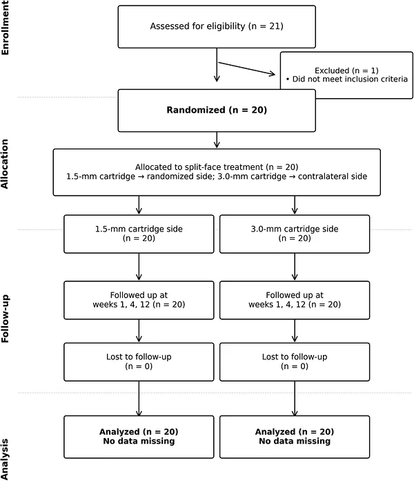
FIGURE 1 CONSORT‐style flow diagram of participant enrolment, allocation, follow‐up, and analysis. Twenty‐one individuals were assessed for eligibility, of whom one was excluded for not meeting the inclusion criteria. The remaining 20 participants were randomized and underwent split‐face treatment, with one forehead receiving the 1.5‐mm cartridge and the contralateral side receiving the 3.0‐mm cartridge. All 20 participants completed all scheduled follow‐up visits at weeks 1, 4, and 12 without losses to follow‐up or missing data. All 20 participants were included in the final analysis.CONSORT 式流程图,展示受试者筛选、随机分配与随访:21 人接受评估,1 人因不符合标准被排除,剩余 20 人随机接受 split-face 治疗并完成第 1、4、12 周全部随访,全部纳入最终分析。2.2
A split‐face design was used in which each participant received the 1.5‐mm cartridge on one side of the forehead and the 3.0‐mm cartridge on the contralateral side, enabling direct within‐subject comparison (Figure 2 ). The side (left or right forehead) receiving each cartridge depth was determined using a computer‐generated random number sequence with a 1:1 allocation ratio. The randomization sequence was generated by an independent investigator who was not involved in treatment delivery or outcome assessment. Owing to the nature of the intervention, the treating dermatologist could not be blinded to cartridge identity. However, participants were unaware of which side received each cartridge depth (subject‐blinded design), and outcome assessors remained blinded to side allocation throughout photographic evaluation.

FIGURE 2 Split‐face study design and randomized cartridge allocation. Representative photographs of the forehead illustrating the split‐face design used in this study. Each participant received the 1.5‐mm cartridge on one side of the forehead (orange ellipse) and the 3.0‐mm cartridge on the contralateral side (blue ellipse).示意 split-face 研究设计与随机 cartridge 分配,展示额部一侧使用 1.5 mm cartridge(橙色椭圆)、对侧使用 3.0 mm cartridge(蓝色椭圆)的代表性照片。2.3
Treatment was performed in a single session using a focused ultrasound device (LIPOGE‐X [ULTLINE]; Eunsung Global Co., Ltd., Wonju, South Korea) equipped with two interchangeable cartridges: 1.5‐mm (0.25 J/dot; 15 dots/shot; 60 shots; total energy, 225 J) and 3.0‐mm (0.25 J/dot; 15 dots/shot; 60 shots; total energy, 225 J) focal depth cartridges. Each treatment line was 25 mm in length, with 5‐mm spacing between adjacent lines. All treatments were performed by a single experienced dermatologist following a standardized protocol. No topical anesthesia or skin cooling was applied.
2.4
Standardized frontal photographs were obtained at baseline, immediately after treatment, and at weeks 1, 4, and 12 after treatment. All photographs were acquired under standardized conditions using a Canon EOS 850D digital camera (50‐mm focal length) mounted on a fixed tripod. Participants were seated on a fixed chair with the head stabilized using a chin rest, maintaining an upright posture with the eyes in primary gaze and a relaxed neutral facial expression to minimize frontalis muscle activity. The camera was positioned 50 cm from the glabella at a constant height, and photographs were obtained against a black background under standardized fluorescent lighting. Three photographs were acquired at each visit, and the same acquisition protocol was applied throughout the study.
Average and maximum eyebrow height (AEH and MEH, respectively) were measured using Image‐Pro Plus software (version 10, Media Cybernetics, USA) by a single blinded assessor at an independent image analysis center (HLK Clinical Center), following a predefined, written measurement protocol. At each visit, three photographs were acquired, and the photograph with optimal focus, complete eye opening, and neutral head position was selected for analysis according to prespecified quality criteria; the remaining photographs served as backups in case of motion blur or suboptimal positioning. A 3‐cm ruler sticker attached to the left zygomatic area served as an internal calibration reference for pixel‐to‐distance calibration independently for each analyzed photograph, correcting for minor variation in camera‐to‐subject distance across visits. With the face viewed frontally, a horizontal reference line connecting the two medial canthi was drawn and extended laterally across the eyebrow region. Five reference points were marked along this line at 8‐mm intervals, starting from the medial side, and the vertical distance from each point to the superior margin of the eyebrow was measured; AEH was defined as the mean of these five distances. MEH was defined as the maximum vertical distance from the same horizontal reference line to the highest point of the eyebrow. The complete photographic documentation and eyebrow height measurement procedure, excerpted from the study protocol, is provided in File S1 .
Additional outcomes included participant satisfaction assessed using a 5‐point Likert scale, treatment‐related pain measured with a 10‐cm visual analog scale (VAS), and monitoring of adverse events throughout the study period. In accordance with the anticipated adverse events specified in the study protocol, the treatment site was also examined at each visit for erythema, edema, tenderness and bruising.
Safety was assessed at each scheduled visit through vital signs (blood pressure, heart rate), physical examination, and urinalysis. Adverse events were recorded throughout the study period using both spontaneous reports and structured questioning.
2.5
Statistical analyses were performed using IBM SPSS Statistics (version 27.0) and R (version 4.4.2). The primary efficacy endpoint was the within‐cartridge change from baseline in average eyebrow height (AEH) at week 12, evaluated separately for each cartridge depth; the within‐cartridge change from baseline in maximum eyebrow height (MEH) at week 12 was a secondary efficacy endpoint. Because the protocol‐defined primary endpoint was evaluated separately for the two cartridge depths, the primary analysis comprised two cartridge‐specific AEH hypotheses (AEH with the 1.5‐mm cartridge and AEH with the 3.0‐mm cartridge). Because two primary hypotheses were tested, Bonferroni correction was applied to the protocol‐specified one‐sided 97.5% confidence interval (CI), yielding a corrected one‐sided 98.75% CI ( α * = 0.025/2 = 0.0125) for each cartridge‐specific AEH analysis, so that the one‐sided family‐wise type I error rate of the primary family was controlled at 0.025. The primary efficacy criterion for a given cartridge was met when the lower bound of the corresponding 98.75% one‐sided CI for the within‐cartridge AEH change from baseline exceeded 0. The two cartridge‐specific MEH hypotheses were handled as a separate secondary family in a serial gatekeeping sequence: they were assessed only after both primary AEH hypotheses had met the Bonferroni‐adjusted criterion and, conditional on passage of this gate, were themselves Bonferroni‐adjusted in the same manner (one‐sided 98.75% CI) and evaluated against the same criterion. The MEH results are reported as supportive and were not used to determine primary efficacy. The within‐subject between‐cartridge comparison was not among the protocol‐specified endpoints and was conducted as a post hoc exploratory analysis; it was not used to determine primary efficacy, no multiplicity adjustment was applied to it, and its results are reported as hypothesis‐generating. No minimal clinically important between‐cartridge difference was prespecified; accordingly, the study was not designed to establish equivalence or noninferiority between the two focal depths. No formal a priori sample‐size or power calculation was performed for the exploratory between‐cartridge comparison; the overall sample size was determined based on feasibility for the within‐cartridge efficacy assessments. Normality of paired differences was assessed using the Shapiro–Wilk test. Paired t ‐tests were used when normality was confirmed, whereas Wilcoxon signed‐rank tests were used otherwise. Within‐cartridge changes from baseline were evaluated separately at each time point using these paired tests, and the resulting time‐point p ‐values are nominal and unadjusted. In addition, in accordance with the protocol‐specified analysis of the secondary time‐course endpoints, the overall effect of time within each cartridge was examined for AEH and MEH with a one‐way repeated‐measures analysis of variance (ANOVA), followed by Bonferroni‐adjusted post hoc pairwise comparisons among the five assessment time points; these analyses are reported as supportive. No data imputation was required because all 20 participants completed every visit.
3
3.1
The study population included 12 women (60%) and 8 men (40%), with a mean age of 33.0 ± 7.0 (range, 25–52) years. Mean height, weight, and BMI were 166.7 ± 8.0 cm, 67.7 ± 15.7 kg, and 24.1 ± 3.9 kg/m 2 , respectively. All 20 participants completed the 12‐week follow‐up without missing data (Table 1 ).
TABLE 1 Baseline participant characteristics (n = 20).
| Characteristic | Value |
|---|---|
| Age, years (mean ± SD) | 33.0 ± 7.0 |
| Range | 25–52 |
| Sex, n (%) | |
| Female | 12 (60%) |
| Male | 8 (40%) |
| Height, cm (mean ± SD) | 166.7 ± 8.0 |
| Weight, kg (mean ± SD) | 67.7 ± 15.7 |
| BMI, kg/m 2 (mean ± SD) | 24.1 ± 3.9 |
| Baseline AEH, mm (mean ± SD) | |
| 1.5‐mm side | 28.71 ± 3.25 |
| 3.0‐mm side | 29.35 ± 2.72 |
| Baseline MEH, mm (mean ± SD) | |
| 1.5‐mm side | 32.89 ± 3.55 |
| 3.0‐mm side | 33.20 ± 3.09 |
3.2
Efficacy results are summarized in Table 2 ; de‐identified individual‐level AEH and MEH values for all 20 participants at every visit are provided in File S2 . In unadjusted within‐cartridge analyses, AEH and MEH significantly increased from baseline at all evaluated time points for both cartridge depths; these time‐point p ‐values are nominal and unadjusted and are distinct from the multiplicity‐corrected week‐12 efficacy assessment reported below. The within‐subject increase was evident immediately after treatment, and mean changes were numerically larger at later visits for selected outcomes; in the supportive repeated‐measures analysis reported below, only the change in AEH between immediately after treatment and week 12 with the 1.5‐mm cartridge differed significantly between follow‐up visits after adjustment.
TABLE 2 Changes in eyebrow height from baseline across all time points.
| n | Post | Week 1 | Week 4 | Week 12 | |
|---|---|---|---|---|---|
| AEH change, mm (mean ± SD) | |||||
| 1.5 mm | 20 | 0.79 ± 0.52† | 1.23 ± 1.34† | 1.52 ± 1.18† | 1.76 ± 1.19† |
| 3.0 mm | 20 | 0.59 ± 0.50† | 0.99 ± 1.22‡ | 1.11 ± 0.99† | 1.23 ± 1.33† |
| MEH change, mm (mean ± SD) | |||||
| 1.5 mm | 20 | 0.59 ± 0.51† | 0.80 ± 1.10‡ | 1.14 ± 1.29† | 1.33 ± 1.40† |
| 3.0 mm | 20 | 0.36 ± 0.56‡ | 0.92 ± 1.57‡ | 0.95 ± 1.49‡ | 0.90 ± 1.80‡ |
With the 1.5‐mm cartridge, the mean AEH change from baseline was 0.79 ± 0.52 mm immediately after treatment and 1.76 ± 1.19 mm at week 12 (all p < 0.001 vs. baseline). With the 3.0‐mm cartridge, the corresponding values were 0.59 ± 0.50 mm immediately post‐treatment and 1.23 ± 1.33 mm at week 12, with nominally significant increases from baseline at all time points (all p < 0.05).
Similarly, the mean MEH change from baseline was 0.59 ± 0.51 mm immediately after treatment and 1.33 ± 1.40 mm at week 12 with the 1.5‐mm cartridge. With the 3.0‐mm cartridge, the corresponding values were 0.36 ± 0.56 mm immediately after treatment and 0.90 ± 1.80 mm at week 12. In these unadjusted analyses, both cartridge depths were associated with within‐subject increases in eyebrow height that remained nominally significant versus baseline at every follow‐up visit (Figure 3 ).
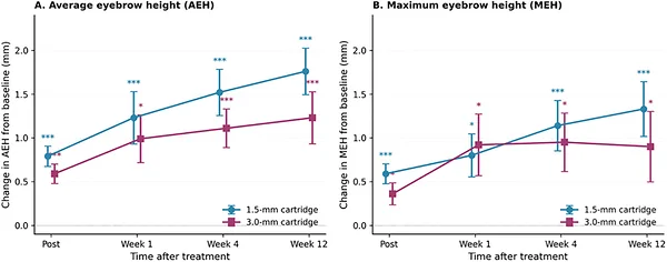
FIGURE 3 Change in average and maximum eyebrow heights from baseline over time for the 1.5‐ and 3.0‐mm cartridges. (A) Change in the average eyebrow height (AEH) and (B) change in the maximum eyebrow height (MEH), expressed as the mean ± 1 SE at indicated time points after a single treatment session. Symbols denote nominal, unadjusted within‐cartridge comparisons with baseline. At week 12, the multiplicity‐corrected primary efficacy criterion for AEH was met with both cartridges; for the secondary MEH endpoint, the separately adjusted criterion was met with the 1.5‐mm cartridge but not with the 3.0‐mm cartridge. *** p < 0.001 versus baseline; * p < 0.05 versus baseline (paired t‐test or Wilcoxon signed‐rank test, as appropriate). n = 20 per side. AEH, average eyebrow height; MEH, maximum eyebrow height; SE, standard error.两种深度 cartridge 的眉高随时间变化折线图(A 平均眉高 AEH、B 最大眉高 MEH,均值±标准误);12 周时两者 AEH 均达到校正后主要疗效标准,MEH 仅 1.5 mm 达到单独校正标准。In the supportive repeated‐measures ANOVA of the within‐cartridge time course, the effect of time was significant for AEH with the 1.5‐mm ( F = 12.60, p < 0.001) and 3.0‐mm ( F = 6.58, p < 0.001) cartridges and for MEH with the 1.5‐mm ( F = 6.31, p = 0.001) and 3.0‐mm ( F = 2.98, p = 0.024) cartridges. In Bonferroni‐adjusted post hoc comparisons among all time points, the comparisons against baseline were consistent with the nominal results for AEH with both cartridges and for MEH with the 1.5‐mm cartridge (adjusted p ≤ 0.041), whereas for MEH with the 3.0‐mm cartridge no comparison against baseline remained significant after adjustment (adjusted p ≥ 0.102). Among comparisons between follow‐up visits, only the increase in AEH from immediately after treatment to week 12 with the 1.5‐mm cartridge was significant (adjusted p = 0.009); no other comparison between follow‐up visits was significant for either outcome or cartridge (adjusted p ≥ 0.240).
At week 12, the lower bound of the Bonferroni‐corrected one‐sided 98.75% CI for the change from baseline in AEH exceeded 0 with both cartridges (1.5‐mm: lower bound, 1.10 mm; 3.0‐mm: 0.51 mm); thus, both cartridge‐specific AEH analyses met the multiplicity‐corrected primary efficacy criterion. For the secondary MEH endpoint, assessed after both primary AEH hypotheses had met the corrected criterion and adjusted separately using the same Bonferroni procedure, the lower bound of the one‐sided 98.75% CI exceeded 0 with the 1.5‐mm cartridge (0.57 mm). With the 3.0‐mm cartridge, although the mean MEH increase remained nominally significant in the unadjusted analysis (0.90 ± 1.80 mm; p = 0.037), the lower bound was −0.08 mm; the multiplicity‐adjusted criterion for the secondary MEH endpoint was therefore met with the 1.5‐mm cartridge but not with the 3.0‐mm cartridge.
3.3
The within‐subject comparison between cartridge depths was performed as a post hoc exploratory analysis; results are shown in Table 3 and Figure 4 . At week 12, the 1.5‐mm cartridge led to a numerically greater AEH increase than the 3.0‐mm cartridge (mean difference, 0.53 ± 1.43 mm; 95% CI, −0.14 to 1.20; p = 0.116). This numerical trend was consistent across all time points; however, none reached statistical significance. For MEH, the 1.5‐mm cartridge showed a mean advantage of 0.42 ± 2.04 mm at week 12 (95% CI, −0.53 to 1.38; p = 0.365). The wide confidence intervals for both the AEH and MEH comparisons indicate that the between‐cartridge differences were not precisely estimated; the nonsignificant p ‐values therefore do not demonstrate equivalence and may reflect limited statistical power. Because no clinically meaningful between‐cartridge difference or decision margin was prespecified, these exploratory results cannot establish equivalence, noninferiority, or a clinically meaningful absence of difference.
TABLE 3 Exploratory within‐subject comparison of 1.5‐ versus 3.0‐mm cartridge (paired differences).
| 1.5 mm | 3.0 mm | Difference | 95% CI | p | |
|---|---|---|---|---|---|
| AEH | |||||
| Post | 0.79 ± 0.52 | 0.59 ± 0.50 | 0.21 ± 0.64 | −0.09, 0.50 | 0.167 |
| Week 1 | 1.23 ± 1.34 | 0.99 ± 1.22 | 0.24 ± 1.36 | −0.40, 0.87 | 0.449 |
| Week 4 | 1.52 ± 1.18 | 1.11 ± 0.99 | 0.41 ± 1.35 | −0.22, 1.04 | 0.341 * |
| Week 12 | 1.76 ± 1.19 | 1.23 ± 1.33 | 0.53 ± 1.43 | −0.14, 1.20 | 0.116 |
| MEH | |||||
| Post | 0.59 ± 0.51 | 0.36 ± 0.56 | 0.23 ± 0.76 | −0.13, 0.59 | 0.191 |
| Week 1 | 0.80 ± 1.10 | 0.92 ± 1.57 | −0.12 ± 1.47 | −0.81, 0.57 | 0.845* |
| Week 4 | 1.14 ± 1.29 | 0.95 ± 1.49 | 0.19 ± 1.45 | −0.49, 0.87 | 0.566 |
| Week 12 | 1.33 ± 1.40 | 0.90 ± 1.80 | 0.42 ± 2.04 | −0.53, 1.38 | 0.365 |
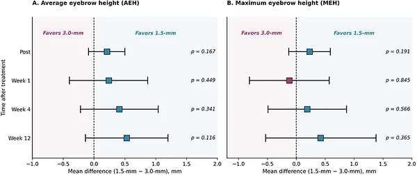
FIGURE 4 Exploratory within‐subject comparison of eyebrow height change between the 1.5‐ and 3.0‐mm cartridges. Forest plot of within‐subject mean differences (1.5‐mm change − 3.0‐mm change) in the (A) average eyebrow height (AEH) and (B) maximum eyebrow height (MEH) at the indicated time points. Squares represent point estimates of the mean within‐subject difference, and horizontal bars represent the 95 % confidence interval. The dashed vertical line indicates zero difference; values to the right favor the 1.5‐mm cartridge, whereas values to the left favor the 3.0‐mm cartridge. In this post hoc exploratory analysis, no between‐cartridge comparison reached statistical significance; the largest numerical mean difference favoring the 1.5‐mm cartridge was observed at week 12 for AEH (mean difference, 0.53 mm; 95% CI, −0.14 to 1.20). p‐values were derived from paired t‐tests (or Wilcoxon signed‐rank tests when the normality assumption was not met). AEH, average eyebrow height; CI, confidence interval; MEH, maximum eyebrow height.深度间比较的森林图,展示各时点 1.5 mm 减 3.0 mm 的组内平均差异及 95% CI;该事后探索性分析中无组间比较达统计学显著性,12 周 AEH 最大数值差异为 0.53 mm(95% CI -0.14 至 1.20)。3.4
Participant satisfaction remained consistently high across all time points. Mean overall satisfaction scores ranged from 4.45 ± 0.51 to 4.60 ± 0.60 on the 5‐point scale. All satisfaction subcategories (procedure, perceived effect, outcome) exceeded 4.0 at every visit.
Safety assessments, including vital signs, physical examinations, and urinalysis, remained within normal limits throughout the study. Pain was mild and transient, with a mean VAS score of 2.20 ± 1.61 immediately after treatment, which decreased to 0 at all subsequent visits. The anticipated adverse events specified in the protocol—erythema, edema, tenderness, and bruising—were not observed in any participant at any visit. No other adverse events, device‐related adverse events, serious adverse events, or unanticipated adverse events were reported during the study period. Given the limited sample size ( n = 20) and the 12‐week observation period, these safety findings should be interpreted as supportive of short‐term tolerability in this cohort rather than as definitive evidence of long‐term safety.
4
In this prospective randomized split‐face pilot study, a single session of focused ultrasound treatment using 1.5‐ and 3.0‐mm focal depth cartridges was associated with statistically significant within‐subject increases in photographically measured eyebrow height sustained through 12 weeks, with high participant satisfaction and no treatment‐related adverse events during the 12‐week observation period in this small cohort. The multiplicity‐corrected primary efficacy criterion for AEH was met with both cartridges, whereas in the separately adjusted secondary MEH analysis the criterion was met with the 1.5‐mm cartridge but not with the 3.0‐mm cartridge. Because the study lacked a sham or untreated control, these within‐subject changes cannot be attributed definitively to the treatment itself.
The magnitude of eyebrow elevation observed in this study was generally consistent with those reported in previous HIFU studies. Oh et al. [ 1 ]. reported a 1.56 ± 0.30 mm mean AEH increase at 12 weeks with 3.0‐mm cartridges in 30 Asian participants. Alam et al. [ 5 ]. reported mean increases of 1.7 mm in average eyebrow height and 1.9 mm in maximum eyebrow height at day 90 using 3.0‐ and 4.5‐mm focal‐depth probes. Choi et al. [ 8 ]. reported a 1.57‐mm AEH increase at week 16 using micro‐focused ultrasound (MFU) at 2.0‐, 3.0‐, and 4.5‐mm focal depths in 38 participants. Moreover, Choi et al. [ 6 ]. reported a 0.91 ± 1.10‐mm AEH increase at week 16. Chen et al. [ 10 ]. reported median eyebrow elevations of 2.16 ± 0.63 mm at 90 days and 1.93 ± 0.57 mm at 180 days using MFU at focal depths of 2.0 and 3.0 mm, indicating sustained yet marginally reduced long‐term effects. Wanitphakdeedecha et al. [ 11 ]. reported an average eyebrow elevation of 1.22 mm at 6 months using macro‐focused ultrasound. In addition, Lio et al. [ 12 ]. reported an approximately 2° increase in eyebrow peak angle, together with increases of 0.89 mm in eyebrow–orbital length and 0.82 mm in vertical palpebral fissure length at day 90. In the present study, AEH increases of 1.76 mm (1.5‐mm depth) and 1.23 mm (3.0‐mm depth) are consistent with these findings and fall within the range of effects reported for HIFU eyebrow lifting (Table 4 ). Table 4 also summarizes the mean or median participant age of each study; among the trials reporting this information, values ranged from 35.4 to 49.8 years, with the youngest comparable Asian cohort reported by Wanitphakdeedecha et al. [ 11 ]. (35.4 ± 6.3 years). The present cohort (33.0 ± 7.0 years) therefore lies at the younger end of this range, although the difference from the youngest of these cohorts is small and the age distributions overlap substantially. These comparisons should be interpreted with caution, as the cited studies differed in study population, device type, treatment protocol, outcome definitions, and follow‐up duration, and therefore do not constitute a direct head‐to‐head comparison of device efficacy.
TABLE 4 Comparison with previously published HIFU studies on eyebrow lifting.
| Study | Sample size (study population) | Frequency (MHz) | Focal depth (mm) | Treatment area | Follow‐up period | Eyebrow elevation | Mean/Median Age (years) | | --- | --- | --- | --- | --- | --- | --- | --- | | Oh et al. [ 1 ]. | 30/Asian | 7.0 | 3.0 | Forehead | 12 weeks | 1.56±0.30 mm (AEH, mean) | NR | | Alam et al. [ 5 ]. | 35/ethnicity NR | 4.0, 7.0 | 3.0, 4.5 | Full face and neck | 90 days | 1.7 mm (AEH, Mean) | 44 (median) | | Choi et al. [ 6 ]. | 22/Asian | 7.0 | 2.0, 2.5, 3.0 | Upper brow, crow's feet, and infraorbital | 16 weeks | 0.91 ± 1.10 mm (AEH, mean) | 48.27 ± 5.44 | | Choi et al. [ 8 ]. | 38/Asian | 4.0, 7.0 | 2.0, 3.0, 4.5 | Forehead | 16 weeks | 1.57 mm (AEH, Mean) | NR | | Chen et al. [ 10 ]. | 40/Asian | NR | 2.0, 3.0 | Upper facial region | 90 d, 180 d | 2.16 ± 0.63 mm (90 d); 1.93 ± 0.57 mm (180 d) (median brow height) | 41.0 | | Wanitphakdeedecha et al. [ 11 ]. | 27/Asian | 4.0 | 2.0 | Forehead, lateral and under eye | 6 months | 1.22 mm (AEH, mean) | 35.41 ± 6.31 | | Lio et al. [ 12 ]. | 12/Asian | 2.0, 4.0, 5.5 | 2.0, 4.5, 6.0 | Forehead, anterior and posterior temporal regions, zygomatic arches | 90 d | +2° (eyebrow peak angle); +0.89 mm (eyebrow–orbital length); +0.82 mm (vertical palpebral fissure length) | 49.75 (median) | | Present study | 20/Asian | 2.0 | 1.5, 3.0 | Forehead | 12 weeks | 1.76/1.23 mm (AEH, mean) | 33.0 ± 7.0 |
Although the device operates at a relatively lower frequency (2 MHz) than conventional MFU systems (4–7 MHz), a parameter that influences tissue penetration depth [ 13 , 14 ], the observed eyebrow elevation fell within the range reported in previous HIFU studies. Because device‐specific acoustic‐field characterization, dosimetry, tissue imaging, and histologic assessments were not performed, the numerically greater elevation observed with the 1.5‐mm cartridge relative to the 3.0‐mm cartridge cannot be attributed to a specific tissue‐level or depth‐dependent mechanism, and any such explanation remains an untested hypothesis.
In the within‐subject exploratory comparison, the 1.5‐mm cartridge consistently showed numerically greater AEH increases than the 3.0‐mm cartridge, although these differences were not significant ( p = 0.116 at week 12). Because this within‐subject between‐cartridge comparison was a post hoc exploratory analysis that was not powered a priori to detect between‐depth differences, the absence of statistical significance should not be interpreted as evidence that the two focal depths produce equivalent outcomes. The 95% CI for the week‐12 AEH difference (−0.14 to 1.20 mm) is relatively wide; with 20 participants, the present data cannot exclude a between‐cartridge difference of a magnitude that would be clinically relevant in eyebrow lifting, where reported treatment effects are themselves on the order of 1–2 mm. Because the numerical trend favored the 1.5‐mm cartridge at every time point for AEH, a type II error remains a plausible explanation for the nonsignificant result. These findings should therefore be regarded as inconclusive and hypothesis‐generating rather than confirmatory and were not used to determine the primary efficacy of either cartridge. Because no clinically meaningful between‐depth difference was prespecified, these data cannot support equivalence, noninferiority, or a clinically meaningful absence of difference between the cartridges; given the absence of formal measurement‐reliability data, the small numerical differences between cartridges should also be interpreted with particular restraint. In the primary efficacy analysis, the multiplicity‐corrected AEH criterion was met with both cartridges, whereas in the separately adjusted secondary MEH analysis it was met only with the 1.5‐mm cartridge; safety profiles were comparable. Whether one focal depth offers a true clinical advantage over the other remains to be determined by adequately powered future studies. Until such data are available, cartridge selection in clinical practice should be guided cautiously by anatomical features, patient comfort, and operator preference, rather than by an assumption of interchangeable efficacy. Future adequately powered studies are needed to determine whether focal depth meaningfully influences eyebrow‐lifting outcomes and to investigate combination strategies that sequentially or concurrently target both depths.
This study had several limitations to consider when interpreting the findings. First, the active‐comparator split‐face design without a sham or untreated control group prevented definitive attribution of the observed effects to the intervention alone, as placebo effects and natural variation cannot be excluded. Second, the small sample size ( n = 20) limited the robustness of the findings and may have underpowered the within‐subject cartridge depth comparison, increasing the possibility of a type II error. Third, although eyebrow height measurements were obtained using a standardized photographic protocol with per‐image pixel calibration and blinded centralized image analysis, all measurements were performed by a single assessor, and no inter‐rater or intra‐rater reliability testing, repeated landmarking, repeat image analysis, or estimation of absolute measurement error was performed. The week‐12 between‐cartridge differences were small (0.53 mm for AEH and 0.42 mm for MEH) and may fall within unquantified measurement variability; they should therefore not be interpreted as evidence of biological or clinical superiority of either focal depth, and the conclusions regarding small between‐cartridge differences have been correspondingly restrained. Future studies should incorporate blinded repeat image reads, intraclass correlation coefficients, and estimates of absolute measurement error to establish the precision of this method relative to the small effect sizes typical of eyebrow‐lifting studies.
Fourth, the 12‐week follow‐up reflected only part of the expected collagen remodeling time course, as HIFU‐induced neocollagenesis typically peaks between 90 and 180 days post‐treatment. The assessment schedule (immediately post‐procedure and at 4 and 12 weeks) complied with the clinical‐evaluation timepoints specified in the approval review guideline for focused ultrasound stimulation systems issued by the Korean Ministry of Food and Drug Safety (MFDS), the relevant chapter of which is reproduced in File S3 . However, regulatory compliance with these assessment timepoints does not in itself demonstrate that 12 weeks is scientifically sufficient to capture the full biological remodeling response or the long‐term durability of the effect; the present results therefore characterize only the early portion of the treatment response, and longer‐term follow‐up studies are warranted. Fifth, the single‐center design and employment of a single treating dermatologist may limit the generalizability of the findings because of site‐specific factors, such as operator technique and patient demographics.
Sixth, the study population consisted of relatively young Korean adults (mean age, 33 years), most of whom had mild eyebrow laxity. This reflects a broader clinical trend toward younger individuals seeking noninvasive HIFU lifting for prevention and subtle refinement, and the cohort's age overlaps substantially with previously published HIFU eyebrow‐lifting trials (Table 4 ). Therefore, the findings may not be directly generalizable to older individuals with clinically significant brow ptosis or more advanced upper facial laxity. Moreover, a 1–2 mm change in eyebrow height may carry a different clinical significance in younger adults seeking subtle refinement than in older individuals with true brow descent. Furthermore, because all participants were Korean, ethnic differences in eyebrow anatomy and soft‐tissue characteristics may further limit extrapolation of these findings to other populations. Finally, this study was not prospectively registered in a public clinical trial registry, which is a limitation with respect to current reporting standards for randomized clinical studies.
5
In this small, uncontrolled pilot study, a single focused ultrasound treatment session using either 1.5‐ or 3.0‐mm cartridges was associated with statistically significant within‐subject increases in photographically measured eyebrow height sustained over 12 weeks. The multiplicity‐corrected primary efficacy criterion for AEH was met with both cartridges; in the secondary MEH analysis, the separately adjusted criterion was met with the 1.5‐mm cartridge but not with the 3.0‐mm cartridge. Both treatments were associated with high participant satisfaction, and no treatment‐related adverse events were observed during the 12‐week study period, which supports short‐term tolerability in this cohort rather than definitive safety. Because this active‐comparator split‐face design lacked a sham or untreated control, these findings support promising within‐subject improvement rather than definitive causal efficacy. The post hoc exploratory within‐subject comparison between cartridge depths did not show a statistically significant difference; this finding should be regarded as inconclusive, as the study was not prospectively powered for this between‐cartridge comparison, and a type II error cannot be excluded. These findings should not be extrapolated beyond the studied population of young Korean adults without further investigation. Larger sham‐controlled studies with longer follow‐up are warranted to confirm these findings, to determine whether focal depth meaningfully influences treatment outcomes across age and ethnic groups, and to assess long‐term treatment durability.
This study received no external grant funding from public, commercial, or not‐for‐profit agencies. The study device and treatment cartridges were provided by Eunsung Global Co., Ltd. This in‐kind support is described in the conflicts of interest statement.
The study protocol was approved by the Institutional Review Board of Chung‐Ang University Gwangmyeong Hospital (IRB No.: 2406‐167‐079). Written informed consent was obtained from all participants. This study was not prospectively registered in a public clinical trial registry prior to participant enrollment.
Hyewon Song and Yuhwa Choi are employees of Eunsung Global Co., Ltd. (Wonju, South Korea), the manufacturer of the focused ultrasound device evaluated in this study. Kwang Ho Yoo is an independent academic clinician affiliated with Chung‐Ang University Gwangmyeong Hospital and declares no personal financial relationship (e.g., consulting fees, honoraria, or equity) with Eunsung Global Co., Ltd. The study protocol was developed by Eunsung Global Co., Ltd. The company provided the focused ultrasound device, treatment cartridges, and logistical support necessary for the conduct of the study. The manufacturer‐affiliated authors (Hyewon Song and Yuhwa Choi) reviewed and edited the sections of the manuscript describing the device and treatment protocol; critical revision of the manuscript's academic content was led by Kwang Ho Yoo. The manufacturer‐affiliated authors (Hyewon Song and Yuhwa Choi) had no role in the collection or quantitative analysis of the primary efficacy data. Kwang Ho Yoo had full independent oversight of the study design, data analysis, and manuscript content. Photographic measurements were performed at an independent analysis center (HLK Clinical Center) blinded to manufacturer affiliation. All authors had full access to the de‐identified study data, were involved in data interpretation, and approved the final manuscript for submission.
声明:中文精读 · 仅供学术参考。内容来自公开文献检索,不代表本人观点,不构成诊疗建议。 医疗美容需在正规医疗机构由执业医师实施。
基于相同主题推荐 · 继续深挖
前瞻性随机 split-face 研究纳入 20 名韩国成人,单次治疗,一侧用 1.5 mm、对侧用 3.0 mm 聚焦超声(HIFU) cartridges(每侧 225 J)。
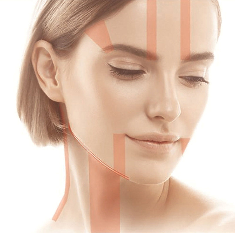
背景:面部老化呈区域特异性凸度增加与沟槽空虚,需分层(浅脂肪层约 3.0 mm、SMAS 约 4.5 mm)而非均一能量;多数既往 HIFU 研究采用全脸均一参数,难兼顾相反容积向
本研究为IRB批准的前瞻性split-face试点研究,纳入3例女性(共6侧面部),一侧注射PDLLA、对侧注射CaHA,于注射后3个月在拉皮术中取对应部位活检行组织学与免疫组化评